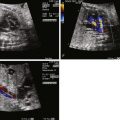
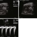
- •
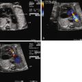
Locate the position of the diverticulum as either at the apex (most common) or originating from the wall of the ventricle.
- •
Determine the presence or absence of a pericardial effusion.
- •
Determine whether there is any ventricular dysfunction or regional wall abnormalities near the site of the “outpouching” because none should be present in a true diverticulum.
- •
Doppler color flow imaging may demonstrate flow between the cavity of the ventricle and the diverticulum.
Anatomy and Anatomical Associations
Outpouchings of the ventricular cavity are referred to as either a “diverticulum” or an “aneurysm” of the ventricle. The terms are frequently used interchangeably; however, some distinctions can be made. A diverticulum is described as having all layers of myocardium within the wall and contracts either normally or near normally, whereas an aneurysm contains components of fibrotic tissue and does not contract normally. A diverticulum is also described as having a narrow neck and is relatively small; aneurysms are more broad-based and can be quite large.
There is an interesting association between a thin-walled ventricular diverticulum and pericardial effusion in the fetus. The pericardial effusion is not related to cardiac dysfunction or hemodynamic abnormality because the diverticulum is often quite small, with no evidence of myocardial compromise. One theory is that the diverticulum acts as a semipermeable membrane allowing for extravasation of fluid into the pericardial space. Intraventricular pressure promotes flux of fluid across and creates a transudate in the pericardial space. These pericardial effusions can become quite large, leading to either cardiac tamponade or pulmonary hypoplasia as it compresses the lungs.
Most commonly, diverticula are seen in isolation. However, they can also be seen in conotruncal anomalies or in association with isolated ventricular septal defects. Diverticulum of the left ventricle can be part of the pentalogy of Cantrell an anomaly consisting of defects in the abdominal wall, sternum, diaphragm, pericardium, and heart. Omphalocele and ventricular diverticulum suggest the presence of this syndrome.
In a review of 35 fetuses with diverticula/aneurysms diagnosed prenatally, 21 were defects in the left ventricle and 14 in the right ventricle. They were located in the apex of the heart in 26, below the atrioventricular valve in 7, and on the lateral free wall in 2.
Frequency, Genetics, and Development
The true incidence of diverticula or aneurysms is not known, because many cases likely are asymptomatic and go unnoticed.
The anatomical features of these outpouchings or “bulges” hint at possible etiologies, although the definitive cause is unknown. Narrow-necked, thin-walled, saccular diverticula are thought to derive from congenital abnormalities of ventricular wall formation and represent a localized, embryogenic defect. Aneurysms appear as broad-based bulges with noncontractile, or dyskinetic, walls and are thought to relate to regional myocardial ischemia or tissue scarring and are perhaps an acquired phenomenon due to disrupted myocardium. These localized areas of ischemia may be due to abnormalities of coronary development in the region affected. Another distinguishing factor is that diverticula typically remain stable in size, whereas ventricular aneurysms can grow, further supporting the notion than the former are discrete congenital abnormalities and the latter relate to ischemic myocardium and respond to intraventricular pressures by potentially changing shape and progressing in size.
Right ventricular aneurysm has been described in the recipient of twin-twin transfusion after fetoscopic laser photocoagulation procedure. The donor died after the procedure and the recipient developed an echogenic focus on the right ventricle free wall with pericardial effusion. Three weeks later, the area developed myocardial disruption with a thin-walled aneurysm and subsequent rupture. The absence of aneurysm before the procedure with identification of an echogenic focus immediately after suggests strongly for an acquired ischemic event, with subsequent myocardial wall scarring and rupture. This case portrays the role of ischemia in the development of fetal ventricular aneurysms, although unlike in this case, most inciting events in other fetuses occur surreptitiously and are of unknown etiology.
Prenatal Physiology
A ventricular diverticulum in the fetus should not cause any hemodynamic compromise on its own; however, when associated with a pericardial effusion, tamponade can occur. The heart can become compressed with impediment to filling, reducing stroke volume. Tachycardia can be seen as the fetal heart attempts to maintain cardiac output. Furthermore, a large pericardial effusion can compress the lungs as well. When long-standing, such compression can impair pulmonary development.
A ventricular aneurysm in the fetus may cause hemodynamic instability, depending upon the size and degree of myocardium affected. If relatively large, contraction mechanics can be negatively affected with overall reduction in cardiac function, poor cardiac output, and development of fetal hydrops. Regions of the myocardium that support valve function can be affected, and these can present with significant tricuspid or mitral regurgitation. Ischemic regions can also be a source for arrhythmia, and ventricular arrhythmias are possible. Thin-walled scarred tissue may also rupture, leading to fetal demise.
Prenatal Management
Echocardiography will demonstrate the architecture of an aneurysm; however, small ventricular diverticula can be difficult to image. A pericardial effusion should prompt a careful search for a diverticulum.
If a large pericardial effusion is present, pericardiocentesis may be indicated. This has been successfully performed relatively early in gestation, even as early as 14 weeks’ gestation. The decision to drain a pericardial effusion will depend upon its impact on hemodynamics or whether there is compression of the lung. Factors that suggest the presence of a hemodynamically significant pericardial effusion include (1) small heart size and general appearance of poorly filled ventricles, (2) single peak filling pattern across the ventricular inflow, (3) Doppler evaluation of the ductus venosus demonstrating reversal with atrial contraction, (4) Doppler evaluation of the umbilical vein demonstrating pulsations, or (5) fetal tachycardia.
Pericardial effusions associated with a ventricular diverticulum can progress and worsen or spontaneously resolve over time. Therefore, if there is a large pericardial effusion but hemodynamics are stable, frequent serial surveillance is important. If a large pericardial effusion is present and the lungs appear compressed, without evidence for any regression of effusion, pericardiocentesis should be considered to relieve intrathoracic pressure and allow for lung expansion.
A ventricular aneurysm should prompt the investigator to look at ventricular function as well as atrioventricular valve regurgitation. Supraventricular arrhythmia, if present, can be treated with maternal administration of digoxin. Ventricular arrhythmias are much more difficult to treat and suggest the presence of “distressed” myocardium, which is an ominous sign.
The fetus with a large ventricular aneurysm and heart failure may benefit from an expectant delivery via cesarean section with an appropriate caretaker team on stand-by, because transitional physiology can add undue load to a sensitive heart, leading to sudden decompensation.
Stay updated, free articles. Join our Telegram channel

Full access? Get Clinical Tree


