Abstract
Background
Abdominal radiographs remain useful in newborns. Given the high radiation sensitivity of this population, it is necessary to optimize acquisition techniques to minimize radiation exposure.
Objective
Evaluate the effects of three additional filtrations on radiation dose and image quality in abdominal X-rays of newborns using an anthropomorphic phantom.
Material and method
Abdominal radiographs of an anthropomorphic newborn phantom were performed using acquisition parameters ranging from 55 to 70 kV and from 0.4 to 2.5 mAs, without and with three different additional filtrations: 0.1 mm copper (Cu) + 1 mm aluminum (Al), 0.2 mm copper + 1 mm aluminum, and 2 mm aluminum. For each X-ray the dose area product (DAP) was measured, the signal-to-noise ratio (SNR) was calculated, and image quality (IQ) was evaluated by two blinded radiologists using the absolute visual grading analysis (VGA) method.
Results
Adding an additional filtration resulted in a significant reduction in DAP, with a decrease of 42% using 2 mm Al filtration, 65% with 0.1 mm Cu + 1 mm Al filtration, and 78% with 0.2 mm Cu + 1 mm Al filtration ( p < 0.01). The addition of 2 mm aluminum filtration does not significantly decrease the SNR ( p = 0.31), CNR ( p = 0.52) or the IQ ( p = 0.12 and 0.401 for reader 1 and 2, respectively). However, adding copper-containing filtration leads to a significant decrease in, SNR, CNR and IQ.
Conclusion
Adding a 2 mm Al additional filtration for abdominal radiographs in newborns can significantly reduce the radiation dose without causing a significant decrease in image quality.
1
Introduction
Although the use of abdominal X-rays is decreasing in adults , they continue to play a crucial role in pediatric imaging, particularly in cases of acute abdomen in newborns . In this population, abdominal X-rays are essential for diagnosing and monitoring intestinal obstruction or necrotizing enterocolitis as they provide evidence of bowel loop distension, pneumatosis intestinalis, or pneumoperitoneum. Despite conventional radiography being known for inducing low levels of radiation, the need for repeated examinations in these situations can lead to a cumulative radiation dose and its impact on human health is still subject to debate .
Moreover, children, especially newborns, appear to have higher radiation sensitivity and are more susceptible to stochastic radiation effects due to their developing bodies and longer life expectancy postexposure . Therefore, optimizing radiation doses and image quality in newborns remains crucial to obtain X-rays with adequate image quality while keeping the radiation dose as low as reasonably achievable (ALARA) yet diagnostically acceptable (ALADA).
One available solution to reduce the radiation dose is the addition of an extra filtration that removes low-energy photons from the beam, as they contribute to the radiation dose without improving global image quality [ , ]. Copper and aluminum are commonly used materials for this purpose in medical imaging. Numerous studies have demonstrated the benefits of additional filtration for thoracic , abdominal X-rays [ , ] and even thoracic CT in adults, as well as for pelvic X-rays in children . In newborns, it has been shown that additional filtration can reduce radiation dose without compromising the quality of thoracic and pelvic X-rays . However, to our knowledge, no study has explored the value of additional filtration for abdominal radiographs in newborns.
Therefore, the aim of this study was to investigate whether the use of additional filtration could reduce the radiation dose of abdominal radiographs in newborns without compromising image quality.
2
Materials and methods
2.1
Phantom
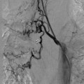
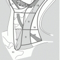
To conduct this study, X-rays were performed using an anthropomorphic newborn phantom, the Kyoto PH-50B Newborn Whole-Body Phantom PBU-80. This phantom accurately simulates the anatomical features of a newborn abdomen, including the spine and pelvic bones, colon, soft tissues, lungs, and mediastinum, which are typically visible on an abdominal X-ray.
2.2
Imaging equipment and acquisition technique
Abdominal radiographs were performed using a Philips DigitalDiagnost X-ray unit, which combines an SRO 33100 ROT 380 X-ray tube with an inherent filtration of 2.5 mm Al/75 Kv and a focal spot (small / large) of 0.6 / 1.2 mm and a maximum voltage of 150 kV. A Trixell DR 35×43 flat panel detector was used. The images were acquired with the anthropomorphic phantom in the antero-posterior lying position, without employing the air gap technique. A source-to-detector distance of 110 cm was used, and the collimation field size at the image receptor was 16 × 15 cm. In accordance with recommendations for pediatric imaging [ , ], no grid was used. All equipment underwent regular quality control tests, and the results were within the specifications provided by the manufacturer.
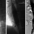
A total of 120 abdominal radiographs were obtained with various additional filtrations available on this device: none, 0.1 mm copper + 1 mm aluminum, 0.2 mm copper + 1 mm aluminum, and 2 mm aluminum ( Fig. 1 ). For each group, 40 radiographs were captured, employing five different kilovoltages (55, 57, 60, 66 and 70 kVp), and for each kilovoltage, eight different tube currents (0.4, 0.63, 0.8, 1, 1.25, 1.6, 2 and 2.5 mAs) were applied. These parameter combinations reflect a wide range of settings commonly used in current practice .

2.3
Radiation dose assessment
The dose area product (DAP) was directly measured by the ionization chamber at the surface of the X-ray tube collimator.
2.4
Signal-to-noise ratio (SNR) and contrast-to-noise ratio (CNR)
To calculate the signal-to-noise ratio (SNR) and the contrast-to-noise ratio (CNR), we measured the mean pixel value (PV) in eight circular regions of interest (ROIs) placed on different structures in the phantom’s abdomen on a dedicated PACS workstation (Carestream Health, Rochester, NY). Six ROIs were distributed along the colon, one on the right iliac bone and one in the left costophrenic recess, to obtain a representative analysis of images while still focusing on the colon, a major organ in the study of abdominal sonography in newborns. The surface area of each ROI was 100 mm². Additionally, a background ROI was drawn within the abdominal soft tissue where there was no overlap of anatomical structures. The ROIs were consistently positioned across all images. The SNR and the CNR were computed using the following equations:
SNR=PViSDbCNR=PVi−PVbσi2+σb22
Related posts:
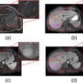 Assessment of a multivariable model using MRI-radiomics, age and sex for the classification of hepatocellular adenoma subtypes
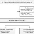 Chest tube placement incidence when using gelatin sponge torpedoes after pulmonary radiofrequency ablation
 Evaluation of a new beads reflux control microcatheter in drug-eluting bead transarterial chemoembolization
 Migrated Superior Vena Cava Stent Repositioned Using a Balloon and Loop Snare Combination
 Bronchial Artery Interventions in Children
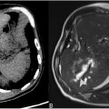
 Carcinomas of the Head and Neck
Stay updated, free articles. Join our Telegram channel

Full access? Get Clinical Tree


