- •
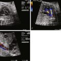
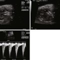
Locate the position of the “echo bright spot” as being solely on the tips of the papillary muscles within the heart.
- •
Search for any additional “echo bright” regions that may suggest an abnormal pathology such as endocardial fibroelastosis (none should be present in the benign form of echogenic foci).
- •
Assess for any atrioventricular valve insufficiency (none should be present in the benign form of echogenic foci).
- •
Assess for ventricular function (should be normal in the presence of the benign form of echogenic foci).
- •
Identify whether there are any structural abnormalities of the heart (none should be present in the benign form of echogenic foci).
Anatomy and Anatomical Associations
Echogenic foci within the fetal heart, or “echo bright spots,” are findings of localized regions of increased ultrasound reflectivity appearing as bright spots within the myocardium. The myocardium itself does not appear distorted by the spot, as might be expected in a tumor or mass, rather the normal myocardium simply appears brighter than its surrounding region. These echogenic foci most commonly appear at the tips of the papillary muscles of the left ventricle. However, they can appear, in order of frequency, within the myocardium of the left ventricle, within the right ventricle, or in both ventricles. They are not associated with any type of structural heart disease and are thought to occur in the normal fetal heart.
The precise pathological origin of this unusual echo-brightness is unclear. Histological analysis of myocardial tissue from high-risk pregnancies in which echogenic foci were noted and termination took place has demonstrated these regions to consist of “mineralization” or regions of calcification with surrounding fibrous tissue. The fetuses analyzed for these studies all had additional anomalies or aneuploidy; hence, it is unclear whether the same histopathology explains the echogenic foci seen in the normal fetus without anomaly or aneuploidy. One alternative hypothesis is that certain myocardial sarcomere genetic allele subtypes, of which there are a number, may phenotypically reflect ultrasound energy to variable degrees. Hence, visualization of an echogenic focus, although not present in all cases, is simply a reflection of a particular normal variant of myocardium that may have these distinctive properties.
Frequency, Genetics, and Development
The precise etiology of these foci is unknown. Studies have demonstrated the presence of echogenic foci within the heart in obstetrical ultrasound of low-risk, midtrimester pregnancy to be at a prevalence of 1.6% to 6.9% of the population. They are typically seen around the middle of the second trimester at approximately 20 weeks’ gestation. Oftentimes, foci disappear upon follow-up evaluation in later gestation.
When first identified in the 1980s, great concern was raised about the possible association of echogenic bright spots within the fetal heart and aneuploidy such as trisomy 21 or association with other genetic syndromes. Multiple large studies have since dispelled this notion. Numerous investigators have concluded that the finding of an echogenic focus is a variant of normal. When there are no other risk factors such advanced maternal age, biochemical abnormalities on screening, or other abnormalities seen on obstetrical ultrasound, no further testing or follow-up is indicated (e.g., amniocentesis is not indicated). The presence of multiple bright spots in both the right and the left ventricles, however, may raise suspicion of aneuploidy and may warrant further analysis. Increased nuchal translucency and bilateral echogenic foci have been reported in a case of 22q11 deletion.
Prenatal Physiology
Although quite common, and not associated with structural heart disease or aneuploidy, do these echogenic foci affect myocardial function in any way? Systolic performance has not been noted to be any different than normal; however, diastolic filling patterns have been reported to be different. In one study, investigators found a decrease in the early diastolic–to–late diastolic phase peak velocity ratio (E : A wave) suggesting greater dependence upon atrial contraction for filling and altered ventricular compliance, although to no clinical significance. A more recent study using sophisticated analysis of myocardial deformation found no differences in systolic or diastolic function in fetuses with echogenic foci in comparison with normal. The issue of whether there might be slight differences in diastolic performance in fetuses with echogenic foci is unsettled; however, if present, these differences do not amount to any clinical significance or clinical manifestations because fetal growth, development, and labor are tolerated as perfectly well as in the normal population.
Prenatal Management
The site and number of foci should be evaluated. Isolated echogenic foci in the papillary muscles of the left ventricle require no further evaluation and are considered normal. Isolated foci to the papillary muscle of the right ventricle are similarly normal. Foci found in both ventricles may require attention. We advocate a high-level obstetrical scan if echogenic foci are found in both ventricles, with consideration of an amniocentesis, in particular, if there are any additional risk factors such as advanced maternal age, a small–for–gestational age fetus, Doppler flow abnormalities in the umbilical artery or ductus venosus, evidence of ventricular size discrepancy, or atrioventricular valve regurgitation.
The presence of echogenic foci may be related to technical considerations related to performance of the ultrasound study. One study reported a much higher read of the presence of echogenic foci when using the apical four-chamber view as opposed to the “lateral” view, or long-axis view, of the heart. Axial resolution is maximized in the apical four-chamber view with the beam of ultrasound running parallel to the papillary muscles, which may explain why the yield was better. Another study highlighted the role of the ultrasonic settings and the operator in the ability to visualize the echogenic foci. These studies suggest that perhaps this particular finding is much more pervasive than it seems, further supporting the notion that this is a common phenomenon seen in the general population, and is visualized depending upon by whom and how the study is performed.
Stay updated, free articles. Join our Telegram channel

Full access? Get Clinical Tree


