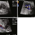
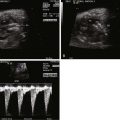
Anatomy and Anatomical Associations
Ectopia cordis is a rare but very dramatic congenital malformation in which the heart is located outside of the confines of the chest cavity. The condition can be classified into four types, depending upon the position of the heart: cervical, thoracic, thoracoabdominal, or abdominal. The most common type is thoracic ectopia in which the heart lies partially or completely outside the chest wall with the ventricular apex pointing cephalad.
Ectopia cordis is commonly associated with other anomalies including intracardiac defects and other “midline” body wall defects. In 1958, Cantrell and coworkers described a “pentalogy” of five associated findings including a midline supraumbilical abdominal wall defect, a defect of the lower part of the sternum, a deficiency of the anterior diaphragm, a pericardial defect at the diaphragmatic surface, and a congenital heart malformation. In this syndrome, the heart disease is often a ventricular septal defect or a conotruncal anomaly such as tetralogy of Fallot or double-outlet right ventricle. Diverticulum of the left ventricle apex is also associated, seen in approximately 20% of cases.
Although Cantrell and coworkers described their particular findings as a constellation, in fact patients may have any one of these alone or a wide spectrum of additional anomalies in association with ectopia cordis. These include abdominal wall defects such as omphalocele, diastasis recti, or gastroschisis as well as craniofacial defects such as cleft lip/palate or neural tube defects. Cases of so-called partial pentalogy of Cantrell have been described, in which there is no sternal defect, but rather a large omphalocele with the heart protruding in a thoracoabdominal manner. Hence, two mechanisms for evisceration of the heart outside of the chest wall have been proposed. One is a defect in the sternum with direct protrusion through, referred to as thoracoschisis. The other is a reverse diaphragmatic hernia in which there is a defect in the diaphragm with the heart protruding into the abdomen in association with an omphalocele within which the abdominal contents including the heart is outside of the body. The embryological mechanisms for these two may be different.
Frequency, Genetics, and Development
Ectopia cordis makes up less than 0.1% of all congenital heart defects. Failure of fusion of the paired cartilage bars of the embryonic sternum leads to a sternal cleft (thoracoschisis), which allows for the potential for the heart to be positioned outside the boundaries of the chest wall. The incidence ranges from 5.5 to 7.9 per 1 million live births. Two thirds of patients are male; one third are born premature or stillborn. Ectopia cordis is part of a developmental field complex defect, which involves deficiency in formation of the sternum, diaphragm, and anterior body wall. A familial pattern has been reported. Ectopia cordis can be associated with a number of genetic and chromosomal anomalies including trisomy 18 and can be experimentally induced in animals through a variety of teratogens.
Prenatal Physiology
The overall physiology and prognosis depend upon the associated anomalies and their severity. Ectopia cordis is commonly part of a pervasive and fundamental abnormality of body wall development. The fetal cardiovascular physiology will be altered based on the type of heart disease seen. One very important manifestation of thoracic and abdominal content evisceration is the development of a small chest and subsequent pulmonary hypoplasia. Although of no physiological consequence in utero during placental support, postnatal survival is limited if pulmonary hypoplasia is present.
Prenatal Management
Careful evaluation of the cardiac position, the amount of heart sitting outside of the chest, the precise type of congenital heart disease, and other associated anomalies is important. Karyotype evaluation is indicated. Oftentimes, suspicion is first raised by the presence of abnormal elevation in first-trimester serum markers, which then leads to early detailed imaging. Ectopia cordis can be diagnosed in the first trimester of pregnancy, as early as 10 weeks’ gestation. Partial protrusion of the heart outside of the chest wall as an isolated finding is unusual, and there are commonly many other associated anomalies. Figuring out the anatomy at the junction of the chest, abdomen, and diaphragm can be a challenge. Fetal echocardiography can be complemented by the use of fetal magnetic resonance imaging to obtain a more precise sense of the anatomy with particular focus on the relationship of the heart to the sternum, diaphragm, and abdominal wall defect. Considering the poor prognosis in most cases, detailed evaluation and counseling are important as early as possible.
Stay updated, free articles. Join our Telegram channel

Full access? Get Clinical Tree


