How to Image the Elbow
- •
Coils and patient position: The elbow typically is scanned with the patient in a supine position with the arm at the side and palm up. A surface coil is imperative for obtaining high-quality images. Occasionally, the size of the patient precludes supine imaging because the surface coil would be too close to the magnet. These patients can be scanned prone with the arm overhead and the elbow as completely extended as possible. Patient comfort is most important for optimal imaging. Positioning the patient comfortably results in a higher yield of images not degraded by motion artifact. Using a vitamin E capsule to mark the area of the patient’s pain or palpable mass is useful for assessing whether the area of concern has been evaluated in the field of view; this is especially important if the examination is normal.
- •
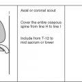
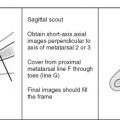
Image orientation ( Box 11-1 ): The elbow should be scanned beginning about 10 cm above the elbow joint through the bicipital tuberosity distally. For convenience, the image should be oriented in the same way as in conventional radiography—the humerus at the top of the image. The axial images should be oriented with the volar surface superiorly.
BOX 11-1
Structures to Evaluate in Different Planes
Axial
- •
Tendons
- •
Annular ligament
- •
Bones
- •
Neurovascular
- •
Muscles
Sagittal
- •
Biceps and triceps tendons (longitudinally)
- •
Anterior and posterior muscle masses
Coronal
- •
Ligaments (medial and lateral)
- •
Medial and lateral muscle masses
- •
Bones (especially medial and lateral epicondyles)
- •
Extensor-supinator and flexor-pronator conjoined tendons longitudinally
- •
- •
Pulse sequence and regions of interest: Axial imaging enables evaluation of tendons, ligaments, bone pathology, and neurovascular bundles. The axial images need to continue through the bicipital (radial) tuberosity to identify the biceps insertion. Coronal imaging is ideal for assessing the integrity of the collateral ligaments and the common flexor and extensor tendon origins. Sagittal images are useful to evaluate the biceps and triceps tendons. Additionally, loose bodies are often best seen on sagittal images. Generally, as with most joint imaging, a slice thickness of 3 mm is reasonable, with a 10% interslice gap (translation 0.3 mm). T1W images show anatomy, particularly in the axial plane, where nerves can be seen surrounded by fat. It is useful to apply fat suppression to T2W fast spin echo imaging because it makes the appearance of pathologic fluid more conspicuous. Because of the unique magnetic susceptibility properties of T2 * imaging, this technique can be used when searching for loose bodies. This technique should not be used in a postsurgical elbow because of the amount of artifact created by micrometallic debris. The degree of artifact surrounding orthopedic hardware is most prominent on T2 * sequences because of the lack of a 180-degree refocusing pulse, and is least prominent on fast spin echo sequences because of the presence of multiple 180-degree pulses.
- •
Contrast: Intravenous gadolinium may provide useful information in the assessment of synovial-based processes or to distinguish cystic from solid masses around the elbow. Intra-articular gadolinium is useful in patients without a joint effusion to detect loose bodies, capsular disruption, and partial tears of the ulnar collateral ligament or to assess the stability of an osteochondral fragment. The dilution is the same for shoulder arthrography (0.1 mL gadolinium mixed with 20-25 mL normal saline). The solution is injected to maximal distention of the joint as indicated by resistance to further injection. This usually occurs at about 10 mL of fluid injected.
Normal and Abnormal
Elbow abnormalities are increasing as the number of individuals participating in weightlifting and throwing and racquet sports increases. (Even couch potatoes are at risk from overuse of the elbow in consuming 12-oz. beverages.) The elbow is one of the most fascinating joints in the body to evaluate. It is the second most congruous joint in the body (knee), and allows for abnormalities to be present in more than one location in the joint. The understanding of elbow pathology is becoming more sophisticated with the advent of improved imaging techniques and the evolution of surface coils. MRI offers superior depiction of muscles, ligaments, and tendons, and the ability to visualize directly bone marrow, articular cartilage, and neurovascular structures.
BONES
Normal Relationships
The osseous anatomy of the elbow allows for two complex motions: flexion-extension and pronation-supination. The elbow is composed of three articulations contained within a common joint cavity. The radius articulates with the capitellum allowing for pronation and supination, and the ulna articulates with the trochlea of the humerus in a hinge fashion. The proximal radioulnar joint is composed of the radial head, which rotates within the radial (sigmoid) notch of the ulna, allowing supination and pronation. The radioulnar joint space also is responsible for one third of the stability of the elbow.
Osseous Disorders
Osteochondritis Dissecans and Panner’s Disease ( Box 11-2 ).
Although osteochondritis dissecans can occur in throwers and in nonthrowers, in dominant and in nondominant elbows, and in the capitellum and in the radial head, it tends to occur in the capitellum of the dominant arm in throwers. The exact cause is uncertain, but the leading hypothesis is that the lesion results from a combination of tenuous blood supply to the capitellum and repetitive trauma at the radiocapitellar joint, resulting in bone death.
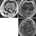
MRI can determine the stability of the osteochondral injury. Unstable lesions are characterized by high signal fluid that encircles the osteochondral fragment on T2W images. Round, cystic lesions may be seen beneath the osteochondral fragment, and abnormal high signal may be seen on the T2W images within the fragment of bone ( Fig. 11-1 ). The overlying cartilage is not always damaged and should be closely inspected. The overlying cartilage is intact in stable lesions and usually is treated with rest and splinting. Unstable lesions are either pinned or excised.

Osteochondritis dissecans has been replaced in the literature by the term osteochondral lesion . This entity should be distinguished from Panner’s disease (an osteochondrosis of the capitellum), which coincidentally also occurs in throwers as a result of trauma. The MRI appearance, patient’s age, and prognosis differ. An osteochondral lesion is seen in slightly older patients (12-16 years), whereas Panner’s disease is in patients 5 to 10 years old. Loose body formation usually is not seen with Panner’s disease, and the entire capitellum generally is abnormal in signal (low signal on T1W images and high signal on T2W images) and may show irregular contour to the capitellum. Subsequent follow-up imaging in Panner’s disease reveals normalization of these changes with little to no residual deformity at the articular surface. An osteochondral lesion can lead to intra-articular loose bodies and significant residual deformity of the capitellum.
A pitfall in diagnosing an osteochondral lesion is the pseudodefect of the capitellum. This pseudodefect occurs because the most posterior portion of the capitellum has an abrupt slope. A coronal image through the posterior capitellum mimics a defect. Examination of this area in another plane and the lack of edema support the pseudodefect as the cause of the irregularity of the capitellum ( Fig. 11-2 ). Additionally, an osteochondral lesion generally begins on the anterior convex margin of the capitellum, whereas a pseudodefect is a finding in the posterior capitellum.

Unstable osteochondral lesions may fragment and migrate throughout the joint as loose bodies. Loose bodies also can occur from purely cartilaginous fragments breaking off in the joint from acute trauma or degenerative joint disease. Loose bodies can become large and cause mechanical symptoms, limiting mobility of the joint, or produce a synovitis that results in an effusion and stiff elbow ( Fig. 11-3 ). Loose bodies may be identified in the posterior compartment in a throwing athlete and are easier to detect on MRI when joint fluid is present in the joint space; they appear as low to intermediate signal structures within high signal fluid. Occasionally, a loose body can have marrow within it. The signal characteristics follow that of fat, high in signal on T1W images. Axial and sagittal images aid in the diagnosis and location of loose bodies. Small fragments of cortical bone result in blooming on T2 * sequences, which may make them more conspicuous than on other imaging sequences.

Fractures.
MRI is useful in evaluating radiographically occult fractures when there is radiographic evidence of a joint effusion, but a fracture is not visualized. Marrow-sensitive sequences (T1W imaging, [fast] STIR, and fatsuppressed fast spin echo) are the most sensitive for assessing the fracture and edema associated with it ( Fig. 11-4 ). Gradient echo is the least sensitive technique for evaluating the marrow. MRI is outstanding for evaluating stress fractures. One type of stress fracture diagnosis that is important in the elbow involves the middle third of the olecranon ( Fig. 11-5 ). This type of fracture is seen in throwing athletes as a result of overload by the triceps mechanism. These fractures can displace and require surgical fixation. Similarly, nonunion of an injury through the olecranon physeal plate can be detected and may require surgical intervention. MRI can adequately assess fractures extending through the cartilage of the physis.


LIGAMENTS
The anterior and posterior portions of the joint capsule are thin. The medial and lateral portions are thickened to form the collateral ligaments. The ligaments of the elbow are divided into the radial and ulnar collateral ligament complexes.
Radial Collateral Ligament Complex ( Box 11-3 )
Normal Radial Collateral Ligaments.
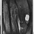
The radial collateral ligament complex provides varus stability. This complex consists of the radial collateral ligament, the annular ligament, the accessory collateral ligament, and the lateral ulnar collateral ligament ( Fig. 11-6 ). The annular ligament surrounds the radial head, and originates and inserts onto the anterior and posterior margins of the lesser sigmoid notch of the ulna. It is the primary stabilizer of the proximal radioulnar joint and is best seen on axial images ( Fig. 11-7 ). The radial collateral ligament proper arises from the anterior margin of the lateral epicondyle and inserts onto the annular ligament and fascia of the supinator muscle ( Fig. 11-8 ). The lateral ulnar collateral ligament is more posterior and is absent in 10% of anatomic specimens; it is thought to provide the primary restraint to varus stress. It is a more superficial and posterior continuation of the radial collateral ligament, arising from the lateral epicondyle and extending along the lateral and posterior aspect of the proximal radius to insert on the ulna at the crista supinatoris ( Fig. 11-9 ).
- •
Restrains varus stress
- •
Lateral ulnar collateral ligament—most important
- •
Radial collateral ligament—less important
- •
Radial collateral ligament complex injury
- •
MRI: increased T2 or complete disruption
- •
Lateral ulnar collateral ligament insufficiency leads to posterolateral rotatory instability
- •
Associated with lateral epicondylosis (tennis elbow)
- •


The origin of the three ligaments is immediately adjacent and deep to the common extensor tendon. Functionally, the lateral ulnar collateral ligament is more important because it is the primary posterolateral elbow stabilizer and maintains the support of the radial head and radioulnar articulation. The radial collateral ligament proper and the lateral ulnar collateral ligament are well seen on coronal images, and both should be evaluated as discrete structures because of their difference in functional significance. The lateral ulnar collateral ligament is often identified on the same coronal image as the pseudodefect.
Abnormal Radial Collateral Ligaments.
Disruption of the lateral collateral ligament complex is more unusual than that of the ulnar collateral ligament complex. Job-related or sports-related injuries usually result in chronic, repetitive microtrauma that produces varus stress. Injury to the radial collateral ligament complex commonly is associated with lateral epicondylar soft tissue degeneration and tearing of the common extensor tendon ( lateral epicondylosis or tennis elbow ). Acute varus injury or elbow dislocation also can be associated with radial collateral ligament complex injury. Insufficiency of the lateral ulnar collateral ligament may result in posterolateral rotatory instability, which allows transient rotatory subluxation of the ulnohumeral joint and secondary subluxation or dislocation of the radiohumeral joint. Rupture of this ligament occurs most commonly as a result of a posterior dislocation or varus stress.
Insufficiency of the lateral ulnar collateral ligament also may occur after a lateral extensor release for tennis elbow (because of the close proximity of the origin of the ligaments and tendons) or with resection of the radial head. This injury most often is seen in patients who sustain a fall on an outstretched hand with resulting hyperextension and varus stress. Laxity of the lateral ulnar collateral ligament after surgical lateral extensor release for tennis elbow has been described as a result of extensive subperiosteal elevation of the common extensor tendon and radial collateral ligament complex during surgery, and as a result of unrecognized lateral ulnar collateral ligament insufficiency preoperatively. Patients frequently complain of locking or snapping of the elbow. The physical examination may produce pain over the lateral aspect of the elbow, a subjective complaint of laxity or instability with varus stress, and a positive lateral pivot shift maneuver.
At surgery, laxity or disruption of the lateral ulnar collateral ligament and the posterolateral portion of the capsule and possible radial collateral ligament laxity can be identified. Reconstruction or reattachment of the lateral ulnar collateral ligament on the lateral epicondyle is performed.
A sprain of the radial collateral ligament complex appears as a thickened or thinned ligament with high signal in and around it. A complete tear shows discontinuous fibers along the radial collateral ligament or lateral ulnar collateral ligament. Proximal detachment or avulsion of their common origin on the lateral epicondyle shows edema and hemorrhage extending into the defect and absence of the fibers of the radial collateral ligament complex ( Figs. 11-10 and 11-11 ). When the lesion is seen in association with lateral epicondylosis, bone marrow edema in the lateral epicondyle and high signal in the extensor tendon group can be identified. If surgical release of the common extensor tendon is being considered, the integrity of the lateral ulnar collateral ligament must be assessed.


Ulnar Collateral Ligament Complex ( Box 11-4 )
Normal Ulnar Collateral Ligaments.
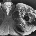
The ulnar collateral ligament complex consists of three bundles: the anterior, posterior, and transverse bundles ( Fig. 11-12 ). The anterior bundle is a thick, discrete ligament with parallel fibers arising from the medial epicondyle and inserting onto the medial coronoid process; it is the most important of the ligaments and is well seen on coronal images. The MRI appearance is that of a low signal linear structure that is flared proximally and tapers distally on all imaging sequences ( Fig. 11-13 ). It is normal to see slight high signal in the proximally flared portion of the anterior bundle. The anterior bundle of the ulnar collateral ligament provides the primary restraint to valgus stress and commonly is damaged secondary to overuse in throwers.
Restrains Valgus Stress
- •
Bundles
- •
Anterior (the important one)
- •
Posterior
- •
Transverse
- •
Best seen on coronal images
- •
- •
Ulnar collateral ligament injury
- •
MRI: increased T2, thickening (partial tear) or complete disruption
- •
- •
Partial ulnar collateral ligament tear
- •
Fluid between distal ligament and ulna (deep fibers disrupted)
- •


The fan-shaped posterior bundle of the ulnar collateral ligament is a thickening of the capsule that is best defined with the elbow flexed at 90 degrees. The transverse bundle of the ulnar collateral ligament is formed from horizontally oriented capsule fibers joining the inferior margins of the anterior and posterior bundles. It stretches between the tip of the olecranon and the coronoid and does not contribute to elbow stability because of ulna origin and insertion. The transverse and posterior bundles are located deep to the ulnar nerve and, in conjunction with the capsule, form the floor of the cubital tunnel. The posterior and transverse bundles are not well depicted on MRI, but their integrity is inferred based on the floor of the cubital tunnel; regardless, they are of limited importance and inconsistently present, and are not considered further here.
Abnormal Ulnar Collateral Ligaments.
Ulnar collateral ligament injury commonly occurs in throwing athletes and may accompany an injury to the common flexor tendon group. Injury to these medial stabilizing structures is caused by chronic microtrauma from repetitive valgus stress during the acceleration phase of throwing.
Complete rupture of the anterior bundle of the ulnar collateral ligament usually occurs suddenly. Patients with acute ulnar collateral ligament ruptures report sudden pain with or without a popping sensation that occurred with throwing, and they are unable to throw after the injury. These injuries are well seen on coronal MRIs. Abnormal signal is identified in the expected location of the linear, low signal structure of the ulnar collateral ligament ( Fig. 11-14 ). The torn fragments also can be identified.

In a large series of throwing athletes, midsubstance ruptures of the anterior bundle of the ulnar collateral ligament accounted for 87% of ulnar collateral ligament tears, whereas distal and proximal avulsions were found in 10% and 3%, respectively. Chronic degeneration of the ulnar collateral ligament is characterized by thickening of the ligament secondary to scarring, often accompanied by foci of calcification or heterotopic bone. Treatment for acute ulnar collateral ligament tears is changing. Conservative treatment is recommended for non-elite athletes (mere mortals) because the flexor-pronator mass keeps the elbow functionally stable, although throwing is limited. Surgical reconstruction for competitive athletes has been recommended for many years.
Partial detachment of the deep undersurface fibers of the anterior bundle of the ulnar collateral ligament also may occur. These patients present with medial elbow pain. The diagnosis with routine MRI is difficult. This type of tear of the ulnar collateral ligament spares the superficial fibers of the anterior bundle and is invisible from an open surgical approach unless the undersurface of the ligament is inspected. These tears are more easily identified after the injection of intra-articular contrast material (MR arthrography). The capsular fibers of the anterior bundle, which normally insert on the medial margin of the coronoid process, show fluid beneath the distal extension of the anterior bundle ( Fig. 11-15 ). This is often a very subtle finding, but can cause functional debilitation in a throwing athlete. Partial tears are treated with repair or reconstruction in athletes.

Synovial Fringe
A slip of tissue extends from posterior to anterior in the lateral aspect of the joint. It is mentioned here in the discussion of the ulnar collateral ligament because with an incompetent ulnar collateral ligament, this shelf of tissue can get impinged in the radiocapitellar portion of the joint. Although the patient may complain of pain on extension after throwing owing to the fringe being pinched, it is the incompetent ulnar collateral ligament that allowed for the development of impingement ( Fig. 11-16 ).