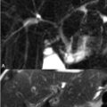
91 Elbow Injuries The annular, lateral ulnar collateral and the radial collateral ligaments comprise the lateral ligamentous structures of the elbow. The normal appearance of the latter is demonstrated on the coronal oblique T1WI of Fig. 91.1A, images acquired in a plane bisecting the epicondyles thus allowing optimal visualization of the elbow’s ligaments and tendons. As illustrated, the radial collateral ligament (black arrow) originates from the lateral humeral epicondyle and joins with the annular ligament surrounding the radius. The common extensor tendon (white arrow), which gives rise to the muscles of hand and wrist extension, also originates from the lateral epicondyle and, as shown in Fig. 91.1A, runs superficial to the radial collateral ligament. In the FS T2WI of Fig. 91.1B, this tendon demonstrates abnormal fluid-like hyperintensity with discontinuity representing a tear of the common extensor tendon origin. Tendinosis with or without a partial tear also demonstrates increased tendinous SI, but does not show a full thickness defect; thickening and thinning of the tendon are most suggestive of tendinosis while focal intratendinous fluid-like signal supports the diagnosis of a partial tear. Full tendinous disruption with intervening hyperintensity on FS T2 or PDWI indicates a complete tear. The radial collateral ligament also demonstrates fluid-like hyperintensity and discontinuity in Fig. 91.1B, consistent with a full-thickness tear. Along with the more posteriorly located lateral ulnar collateral—which transverses the radiocapitellar joint superficial to the annular ligament—the radial collateral ligament prevents posterolateral rotatory elbow instability—a condition leading to perching of the trochlea upon the coronoid, a predisposition to further injury.
Stay updated, free articles. Join our Telegram channel

Full access? Get Clinical Tree


