Fig. 32.1
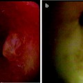
A patient with GOLD III COPD, diabetes, and previous history of CABG and poor vascular condition used aspirin and was presented with hemorrhagic sputum. Superficial spreading squamous carcinoma reconfirmed by a panel of pathologists. Lesion extent was established under autofluorescence bronchoscopy extensive sampling of mucosal biopsies. (a) High-resolution CT scan with 1-mm slice thickness and FDG-PET scan did not show any abnormality; thus, this extensively spreading intraluminal lesion is classified as CT/PET occult. Locally, he was treated with argon plasma coagulation (b), and the necrotic crust can be seen immediately after APC treatment (c). He remains disease-free after 5 years follow-up. Recently, squamous cancer recurrence was diagnosed in the orifice of the superior part left upper lobe bronchus, and due to invisibility of distal tumor extension, external radiation treatment with respiratory gating has been commenced. There is a slight thickening on the HRCT scan seen, and locally FDG-PET scan shows local avidity
APC can be used for burning superficial layer of early-stage mucosal cancer of several millimeter thickness. This is a comparable strategy to using CO2 laser or ultraviolet light excitation of Photofrin II®, to deliberately obtain superficial rather than obtaining too deep necrosis.
Cryotherapy (see separate chapter) has the advantage of preserving bronchial cartilage with less scar tissue formation, which may be important in dealing with segmental and subsegmental location of tumors. However, results are not immediate, and tissue depth effect is difficult to predict while repeated cooling and thawing takes more time. The use of cryoablation has been recently reported in which larger surface area can be cryosprayed much faster.
Brachytherapy (see separate chapter) is a much more complex and expensive facility; special logistics and good collaboration with the radiation oncologist are essential. Even high-dose rate brachytherapy cannot provide immediate solace for emergencies, as several treatment fractions are needed. This is in stark contrast to heat tissue applications such as EC, APC, and lasers which can be obtained in a single treatment session. Therefore, tissue-heating methods, i.e., lasers, EC, or APC, are the only techniques that can provide immediate benefit for rapid recanalization of airway blockage.
Clinical Background
Unfortunately, interventional pulmonologists mostly deal with advanced-stage lung cancer with local tumor growth causing imminent suffocation and respiratory failure. Obstruction may be caused by intraluminal tumor growth as well as extraluminal airway compression by mediastinally located tumor and enlarged lymph node masses adjacent to the tracheobronchial tree, and bronchoscopic debulking or combined with stenting is the only treatment choice.
The majority of patients are usually presented with endstage cancer and comorbidities; hence, their often poor condition and the negative selection of patients are such that morbidity or mortality of bronchoscopic intervention can be quite significant.
With such a clinical presentation, the easy logistics of EC and APC and familiarity of many with electrical appliances make their use for the clinical and outpatient setting more readily accepted.
Palliation and Treatment with Curative Intent
The effectiveness of interventional bronchoscopic treatment for immediate palliation of central airway obstruction has been established, often being the only treatment alternative left. Conceptually, the use of EC and APC is no different than applying Nd:YAG laser as described earlier. Easy logistics allow practical management in the daily practice of a bronchoscopy unit (2–4). As extensive investigations, e.g., CT scan, lung function measurements, and blood gas analysis, prior to intervention in patients with imminent respiratory failures are unfeasible, clinical findings of stridor and severe dyspnea justify immediate action. EC and APC can then be applied more easily for tumor coagulation prior to mechanical debulking and stenting.
EC and APC can better safeguard unexpected bleeding after taking biopsy as increasing number of patients at risk have cardiovascular comorbidities and are routinely taking aspirin and clopidogrel. Many educational workshops are now dealing with competency skill training that better prepare operator and team members to act properly and decisively in case of adverse events during bronchoscopy.
By either using the rigid scope or working through the endotracheal (ET) tube, the interventional pulmonologist can perform tissue coagulation with EC or APC probe and recanalize the central airways more effectively. The rigid scope with its larger working channel obviously better provides access for safer manipulation and primarily in safeguarding ventilation. Despite the wider acceptance of using only devices that suite the flexible bronchoscope, the blocking effect of the flexible bronchoscope can jeopardize safety. The proper execution of any interventional technique depends on the readiness and expertise of the team that is familiar with various procedures, including proficiency in using the rigid instruments. Therefore, one should always realize that technique per se is not the only factor that determines success regardless of the use of intraluminal tumor debulking that is easier to be applied.
Treatment for Intraluminal Non-lung Cancer Lesions
Interventional pulmonologists can also provide alternative treatment strategies after diligent consultation within the thoracic multidisciplinary team, for other intraluminally located tissue abnormalities.
Apart from lung cancer, involvement of central airways by tumor metastasis, benign lesions, and slow-growing lesions such as typical carcinoid, even malignant fibrosarcoma, does warrant a proactive role for the interventional pulmonologists to be involved in the care of these patients. The argument that a potential delay in surgery will jeopardize patients’ outcome has not been proven in our longitudinal study with regard to bronchoscopic treatment of bronchial carcinoid. Given current knowledge on neuroendocrine tumors, this is in retrospect not surprising. Even after years of postponement, surgical resection would not have been different regarding extent of resection or outcome in the several cases that there was residual tumor or local recurrence in the bronchial wall. The various bronchoscopic techniques for benign processes must be seen in the same conceptual line as foreign body removal, in which surgical resection should remain the last resort. Any minimally invasive technique that can be first commenced in trying to solve the problem before performing major surgery is preferable rather than an immediate and hasty surgical approach.
Indeed, disease management has become a concerted effort between various disciplines by fully exploiting the different input from expert team members, including interventional pulmonologists. Increasing understanding about tumor behavior and clonal cell growth and behavior in current era of molecular biology and also in dealing with early-stage nonsmall-cell lung cancer is of paramount importance. The involvement of interventional pulmonologists in pulmonary medicine and medical oncology can be very supportive regarding early detection, staging, and treatment strategies, both for central airway and lung parenchymal abnormalities.
Future Strategies
Current interest in stage shift as the primary goal in a lung cancer screening setting poses a great challenge for its clinical management. As earliest stage cancer, i.e., carcinoma in situ and alveolar adenomatous hyperplasia, involves subcentimeter lesions, relying only to the gold standard of histology classification and surgical resection is currently inappropriate.
One may still argue that nonsurgical approaches are still not acceptable until data from phase III prospective trials have shown similar efficacy. However, the potential values of alternative techniques such as interventional bronchoscopy and radiation therapy for clinically unfit patients have been established.
While attention has been put on screening the population at risk for relatively healthy individuals, we must not forget that the clinical reality of increasing number of ageing patients with comorbidities remains the bulk of our care. This is a great challenge to further explore the many potentials of non- and minimally invasive techniques in better preserving quality of life and in improving the cost efficiency of our medical care system rather than relying on accepted standard diagnostic and therapeutic avenues including major surgical approach.
For relatively healthy individuals with early cancer that can tolerate surgical resection, lead time in carcinogenesis increases the potential long-term effect of overdiagnosis, if combined only with aggressive management that has been implemented at the cost of quality of life.
The low positive predictive value of current diagnostic algorithms such as sputum cytology and low-dose spiral CT despite still requires much improvement. Although CT screening data show that more early-stage cancers are being detected and curatively treated, controversies about overdiagnosis are still heavily debated, and downstream morbidities and mortalities related to early detection programs may become a significant issue. Alternative approaches may significantly reduce downstream morbidities, mortalities, and costs not only in a screening program but also in our daily care of the patients by virtue of advancements of non- and minimally invasive technologies in terms of early detection, accurate minimally invasive staging, and local treatment that is potentially curative, as tumor stage is the most important determinant for cure.
Summary
Minimally invasive techniques in the field of interventional pulmonology have led to a better understanding of thoracic disease processes. Combined with current advances in noninvasive imaging, pathology, molecular biology, medical oncology, and radiation oncology, technical development allows us now to combine all expertise for optimally choosing a tailored and personalized strategy for each patient.
Proper consultations within members of the thoracic oncology and respiratory teams can better suit diagnostic, staging, and treatment incentives with optimal preservation of quality of life of the at-risk individuals involved. Increasingly in the ageing population, many individuals suffer from comorbidities, and a more coherent approach toward disease management is warranted.
Treatment use of electrocautery and argon plasma coagulation is just part of the armamentarium for optimizing our care in the daily routine of a bronchoscopy unit.
The encompassing issues of early detection, staging, tumor biological behavior, and treatment, however, are the important determining factors to be taken into account before making a proper decision about disease management that is aimed for a tailored strategy in each patient.
Suggested Reading
1.
Barlow DE. Endoscopic applications of electrosurgery: a review of basic principles. Gastrointest Endoscop. 1982;14:61–3.
4.
5.
6.
Grund KE, Storek D, Farin G. Endoscopic argon plasma coagulation (APC) first clinical experiences in flexible endoscopy. Endosc Surg Allied Technol. 1994;2:42–6.PubMed
7.
8.
Sutedja G, Bolliger CT. Endobronchial electrocautery and argon plasma coagulation. Interventional bronchoscopy. Prog Respir Res. 2000;30:120–32. Basel Karger.CrossRef