Embolization Agents


Embolization Agents


Charles E. Ray, Jr., McKinley C. Lawson and Jason R. Bauer


The principle of catheter-directed embolization is to acutely stop flow in a blood vessel or vascular territory by way of mechanical occlusion using a temporary or permanent agent. Along with angioplasty, embolization has been a unique defining procedure in the history of interventional radiology. The elegance of the technique underscores the value of minimally invasive therapy. Since the first reported cases using autologous clot in 1972,1 embolization techniques have evolved with the use of many agents and devices to yield truly impressive results. Traumatic injuries resulted in the first common application of embolization techniques, where until the late 1970s surgery was considered the only viable option for suspected vascular injury. This technique has evolved to include nearly every vascular territory and has been used in such diverse clinical applications as treatment of tumors, vascular malformations, varicosities, aneurysms and pseudoaneurysms, fibroids, and gastrointestinal bleeding.


An understanding of the available agents and devices is key to using them appropriately. Device selection is driven by the vascular territory to be embolized, the permanence of occlusion, and the degree of occlusion—proximal or distal—desired. In the past, choices were limited to a basic array of agents including polyvinyl alcohol (PVA) particles, Gelfoam, and coils. Now the armamentarium has expanded. Fundamentally, the techniques are unchanged, but the range of products available requires careful assessment in agent selection.


Embolization procedures have evolved from single large-catheter (7F) proximal embolization to coaxial catheter-guided subsegmental vessel selection. Technologic advancements of embolic agents have led to wider applications of the technique. As microcatheters and wires have evolved, the ability to perform subselective third- and fourth-order angiography not only uncovers many sites of vascular injury or supply that previously went undiagnosed but also allows precise delivery of embolic agents. Similarly, digital subtraction angiography (DSA) has led to improved diagnostic sensitivity, and with the development of roadmapping capabilities, subsequent vessel selection and treatment have also been improved. Changes in practice are nowhere more apparent than in gastrointestinal hemorrhage, where many years ago, coils and particles supplanted vasopressin infusion as the transcatheter treatment of choice.


Regardless of the clinical scenario, the ideal embolic agents are those with easily reproducible deployment and delivery and those that do not obstruct the delivering catheter. Understanding of device size, ongoing coagulopathy, vessel size, consequences of irreversible tissue loss, ease of deployment, rapidity of onset, and likelihood of nontarget embolization are considerations every interventionalist weighs when planning an embolization treatment. No one agent is perfectly suited for every situation; in fact, many agents such as Gelfoam (Pharmacia & Upjohn, Kalamazoo, Mich.) and coils or particles and coils may be used together for best results. Understanding of specific agents will ultimately mitigate unexpected results.


In this chapter we will discuss the types of agents available and their applications resulting from both in vivo behavior and clinical results produced. Special attention will be paid to specific clinical scenarios that are historically challenging for the interventionalist. The discussion of biologically active embolic agents functioning as vehicles for treatment (e.g., radioactive spheres, islet cell transplants) will be reviewed in other chapters.


Types of Agents Used


With the increasing number of embolic agents available to the operator, it is often difficult to determine which type of agent would best be used for any given clinical indication. Generally speaking, agents can be separated into one of four types: permanent large-vessel occluders, permanent small-vessel occluders, temporary large-vessel occluders, and temporary small-vessel occluders. The most commonly used embolic agents, separated into these four categories, are presented in Table 10-1.


When deciding which embolic agent is most appropriate for any given application, the authors pose three questions. Is the vessel to be embolized a large vessel (anything that can be seen angiographically to the level of small to medium arterioles) or a small vessel? Is the desired occlusion permanent or temporary? Is cellular or organ death desired? Once these questions are answered, the choice of embolic agent becomes more obvious (Table e10-1).


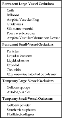
Permanent Large-Vessel Occlusions


Coils


When permanent occlusion of large vessels is desired, coils are most commonly employed for their ease of use and availability. There are many considerations when selecting coils for embolization, beginning with size—the diameter of the wire that forms the scaffold of the coil and the diameter of the unconstrained coil. Coils are offered in a range of diameters from 0.010 to 0.052 inch and are made from steel or platinum, the latter being softer and more radiopaque. Microcoils, which measure 0.018 inch or less, are suitable for deployment through a microcatheter, whereas larger coils are delivered through standard (4F-7F) catheters. One must be careful not to use a microcoil with a standard-sized delivery catheter, since microcoils may coil within the delivery catheter and cause a catheter occlusion. Coils are also available in different geometric designs—Tornado-type coils (Cook Medical, Bloomington, Ind.) taper from base to tip; Interlock-type coils (Boston Scientific, Natick, Mass.) are available in cube, two-dimensional helical, and diamond shapes; and standard coils are untapered and cylindrical. Deployed coil diameters range from a few millimeters to several centimeters. Coils may be bare metal but typically have constituent thrombogenic fibers. Coils are preloaded and configured as a straightened wire in a metal or plastic sleeve, with a pusher to load the coil into the hub end of the delivery catheter.


Coils may be “pushed” coaxially or “chased” with a saline bolus during delivery. The stiff end of the delivery wire is used to partially advance the coil through the proximal portion of the delivery catheter. After the coil is loaded, it is advanced with a floppy-tipped wire. Where less precision is required and there is stable catheter access, a coil may be chased with a saline bolus. “Liquid coils” are ultrasoft platinum coils without attached fibers that are delivered hydrostatically by injection of fluid. In very large vessels that require embolization, occlusion may require a “coil” in the form of a Teflon wrapping from a Bentson wire or wire mandril (discussed later). In all cases, the goal is coil deposition near the end of the catheter, without significant downstream migration.


Strategies for keeping coils in a predictable pack include “scaffolding” by using a larger coil to fix smaller or more flexible coils behind it. Anchoring a coil in a side branch vessel may also facilitate packing. Other techniques include packing coils behind a strut such as an Amplatz vascular occluder device (discussed later) or behind an inferior vena cava (IVC) filter.


Nester coils (Cook Medical) deserve special mention. They are very soft and pliable coils that conform to target vessel anatomy once delivered. They are fibered coils on a platinum strut and are available in both standard and micro sizes. They are commercially available in a wide range of diameters (3-12 mm); however, the diameter is nominal, since the coil “bunches” and forms an unpredictable shape at the target site. The authors use Nester coils only after a predictably shaped coil is first delivered; the initial coil acts as a backstop and precludes a portion of the Nestor coil from extruding out the distal vessel as a “tail.”


Detachable coils offer maximal control, potentially allowing the operator to advance, retract, and reposition the coil before final deployment, thereby affording some measure of safety in high-flow systems where downstream migration can be catastrophic. Pulmonary arteriovenous fistulas are an example where sizing of the first coils is essential. Though conventional coils are simple to use, if the coil is the wrong size or becomes malpositioned during delivery, retrieval is only possible if the coil is still completely within the catheter or accessible by using snare techniques or grasping forceps.


Some detachable coils are released when a weld is broken under pressure or electrolytically. For example, GDC Detachable Coils (Cook Medical) and Matrix Detachable Coils (Boston Scientific) have a connection between the coil and delivery wire that “dissolves” when the operator applies a current to the delivery wire. Others, such as the Interlock-35 System and its derivatives (Boston Scientific), feature a mechanical connection between the coil and delivery wire that disengages when the detachment zone at the base of the coil passes beyond the tip of the delivery catheter. Alternatively, some detachable coils use a release mechanism that relies on fine threads at the base of the coil and at the distal end of the delivery wire (or mandril) that are disengaged by rotating the delivery wire. This system was first described with the Jackson detachable coil2 and has evolved to become the Flipper Delivery System (Cook Medical). It has been argued that this system affords better control, since the coil can be retrieved even when the threads have passed beyond the delivery catheter tip. Many of these detachable coils are now available in 0.035-inch diameter.


Surface-modified coils of the hydrogel type (HydroCoil Embolic System [MicroVention Inc., Tustin, Calif.]) have recently completed evaluation by a randomized controlled trial.3 These coils employ a thin hydrophilic coating that hydrates and expands when placed intravascularly. Compared to traditional platinum coils, this coating (in principle) leads to better aneurysm occlusion/packing; uncoated platinum coils may leave aneurysms only 20%-30% volumetrically occluded with delivered material, versus approximately 70% with the hydrogel system.4 While the remaining void is filled with coil-induced thrombus, this clot remains unorganized for an extended period of time. It is thought that aneurysm regrowth may be related to thrombolysis and recanalization.3,5 Major aneurysm recurrence (incomplete occlusion on follow-up imaging) is estimated to occur in up to 20% of patients at 6 months after treatment with bare platinum coils.3 Through greater volumetric packing, the hydrogel coils are intended to decrease recanalization and aneurysm recurrence. Whether this will ultimately result in improved clinical outcomes is uncertain. The aforementioned randomized controlled trial noted little difference in terms of bleed or re-bleed, but there were fewer angiographic recurrences in the HydroCoil group.3


Hydrogel-coated coils can be delivered through microcatheters. However, since they expand to a diameter several times their initial measurement, they can only be retracted into the microcatheter for a few minutes following contact with blood, reaching maximum diameter approximately 20 minutes after exposure.4 Complication rates appear to be similar between HydroCoils and traditional coils,3,5 though there has been concern regarding a statistically nonsignificant increased occurrence of hydrocephalus.3 Additionally, some authors initially reported increased rates of thromboembolic complications (primarily clot on coil) before systematically adding aspirin to full heparinization during the embolization procedure.5 HydroCoil steaming/softening before deployment has also been necessary to decrease coil stiffness prior to delivery, though newer less stiff coils are now available.5


Newer coils in development are those with biologically active coatings contributing to thrombogenicity or drug delivery, those with nitinol components to maximize stability, and fully retrievable coils to expand the range of temporary embolization options.


Balloons


In the past, balloons were used in different clinical scenarios where a permanent, proximal, large-vessel occlusion was desired. They especially made their mark as occluders during pulmonary arteriovenous malformation (AVM) embolizations.6 In a small percentage of balloons, deflation occurred post embolization. This phenomenon, coupled with the success and ease of use of coils, led to the removal of balloons from the commercial market in the United States.


Amplatzer Vascular Plug


The Amplatzer Vascular Plug I (AVP I [St. Jude Medical, St. Paul, Minn.]) is a device formed from nitinol mesh. The AVP I should be distinguished from the Amplatzer Vascular Occluders, which are a series of biconcave nitinol discs used to percutaneously occlude a septal defect, patent foramen ovale, or patent ductus arteriosus.


The AVP I is a self-expandable device with a postdeployment diameter varying from 4 to 16 mm; the smaller-diameter devices (≤8 mm) are delivered via a 5F guide catheter delivery system, and the larger-diameter devices (≥14 mm) require up to an 8F delivery system. Since the device is made from nitinol, it is relatively radiolucent; the two platinum bands that hold the device together at either end serve as radiopaque markers. The device is secured to the delivery system by a single steel microscrew, and deployment of the device is accomplished by unscrewing the device from the delivery wire. The manufacturer of the device recommends oversizing the device by 30% to 50% (e.g., a 10-12 mm plug would be used for occlusion of an 8-mm vessel). This oversizing allows for precise placement without the risk of distal malposition.


The Amplatzer Vascular Plug II (AVP II [St. Jude Medical]) was introduced several years after the first iteration. It consists of three distinct nitinol meshes connected together as one device. The intent is to provide an increased metal surface area to facilitate quicker and more complete thrombosis. In addition, the AVP II requires less oversizing (20%-30%) compared to the AVP I.


Two other iterations of the AVP are available. The AVP III device is a complex-shaped device intended specifically for use in very high-flow systems. The AVP IV can be delivered via standard 0.038-inch catheter delivery systems and has a reverse hourglass configuration. The AVP IV is currently in the premarket evaluation phase.


Small Intestine Submucosa


The Dotter Interventional Institute and Cook Biotech (Lafayette, Ind.) have teamed to develop small intestine submucosa (SIS) as an embolic agent. Small intestine submucosa is U.S. Food and Drug Administration (FDA) approved as an agent for soft-tissue repair, skin wound dressing, and urethra sling and anal fistula repair, but has not yet been approved for endovascular use.


Described in animal studies as an embolic agent for abdominal aortic aneurysm endoleaks7 and as the membrane for a novel endovascular occluder,8 the material has yet to be used in large published human trials.


The SIS material is composed largely of proteoglycans, collagen, and glycosaminoglycans.7 There are no published reports that describe anything other than a simple mechanical occlusion (e.g., no notable inflammatory response), and the occlusion appears to be permanent (D. Pavcnik, personal communication). Per the investigators, when used, it has a consistency similar to Gelfoam. Detailed description of how to prepare the agent is given elsewhere,7 but presumably, prior to arriving on the market for general use, the process of preparation will become less onerous.


Bare Angiographic Wires


In embolization cases requiring extremely large numbers of coils owing to the large size of the abnormality to be embolized, bare guidewires, or rather their outer Teflon wrappings, can be used as embolic agents. Although these wires have many drawbacks during use, in certain clinical circumstances they may prove to be a viable option.


To use guidewires as embolic agents, the outer Teflon wrapping is used for the embolization, while the inner mandril and metal safety wire connecting both ends of the wire are removed. The easiest way to accomplish this is to cut the outer Teflon sheath and metal stay-wire to the desired size. Once the outer elements are cut and the inner mandril is exposed, the Teflon sheath can be stripped off the mandril by using a hemostat or thumbnail pressure. It should be noted that if the sheath does not come off easily, the inner metal “suture,” or mandril, may not be completely severed. The wire sheath can be loaded into the delivery catheter and pushed out the end hole with a standard (intact) guidewire.


This technique is typically reserved for very large aneurysms/pseudoaneurysms that would otherwise require an inordinate number (dozens) of embolization coils. While the non–fiber-bearing wires would be unlikely to produce significant thrombosis on their own, they are used primarily to occupy volume within the aneurysm sac and, hopefully, decrease the number of additional embolic agents placed for actual thrombosis.


Two caveats should be mentioned. First, when the wire sheaths are deployed, they tend to conform to the outer contour of the aneurysm, essentially “outlining” it. Although not believed to place an undue amount of stress on the outer wall of the sac (owing to the flimsy nature of the sheath), the inner portion of the sac remains relatively unaffected by the embolization materials. Second, once the wire sheaths are deployed, repositioning can be extremely difficult. Snares can be used to remove malpositioned wire sheaths, but the lack of an inner mandril and the “accordion” properties of the sheath cause the snare to essentially pull the proximal end of the sheath without moving the distal end.


Dec 23, 2015 | Posted by in INTERVENTIONAL RADIOLOGY | Comments Off on Embolization Agents

Full access? Get Clinical Tree

Get Clinical Tree app for offline access