KEY FACTS
Terminology
- •
Gas-forming upper UTI involving renal parenchyma &/or perinephric space
Imaging
- •
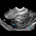
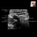
Highly echogenic areas within renal sinus and parenchyma with “dirty” shadowing
- •
Ring-down artifacts: Air bubbles trapped in fluid
- •
Perinephric fluid collections may be seen
- •
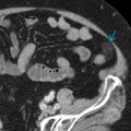
Type I (33%): Parenchymal replacement by gas, ± crescent of subcapsular or perinephric gas
- •
Type II (66%): Renal or perirenal fluid abscesses with bubbly gas pattern ± gas within renal pelvis
- •
Evaluation for psoas abscess and spinal osteomyelitis essential
- •
CT may help further delineate location and extent of renal and perirenal gas
Top Differential Diagnoses
- •
Emphysematous pyelitis
- ○
Gas limited to renal collecting system and pelvis, not parenchyma
- ○
Less clinically serious than emphysematous pyelonephritis, unless obstructed
- ○
Pathology
- •
Single or mixed organism(s) infection
- •
Escherichia coli (68%), Klebsiella pneumoniae (9%)
- •
Proteus mirabilis , Pseudomonas , Enterobacter , Candida , Ilostridia species
Clinical Issues
- •
Extremely ill at presentation: Fever, flank pain, hyperglycemia, acidosis, dehydration, and electrolyte imbalance
Scanning Tips
- •
Gas in perinephric space or perinephric collections may obscure kidney