Endobronchial Tumor
Jud W. Gurney, MD, FACR
Key Facts
Imaging Findings
Air crescent around lesion should suggest endobronchial lesion (also seen with intracavitary lesions)
Bronchus sign: Bronchus leading to peripheral nodule
Lesions have variable density, may contain fat or calcium or low-attenuation material from necrosis
Contrast enhancement: Seen primarily with carcinoid tumors, less commonly mucoepidermoid carcinoma or leiomyoma
Long axis of tumor may parallel course of airway or conform to branching pattern of airways
Iceberg tumors have components both within and external to lumen
Top Differential Diagnoses
Mucus Plugs
Foreign Bodies
Tracheobronchopathia Osteochondroplastica
Broncholith
Pathology
Malignant endobronchial tumors
Non-small cell bronchogenic carcinoma (> 95%)
Carcinoid
Benign endobronchial tumors
Hamartomas (70%)
Clinical Issues
Iceberg tumors cannot be resected bronchoscopically
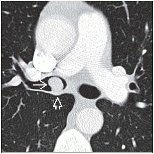 Axial CECT shows a smooth round nodule in the right main bronchus  . The posterior wall is thickened . The posterior wall is thickened  . . |
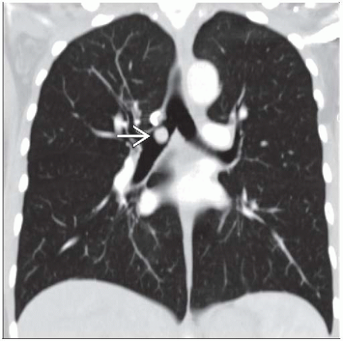 Coronal CECT reconstruction shows a nodule  in the right main bronchus in this patient with a carcinoid tumor. in the right main bronchus in this patient with a carcinoid tumor. |
TERMINOLOGY
Definitions
Hemoptysis: Expectoration of blood from lower airways or lung
Massive hemoptysis: ≥ 600 mL blood/24 hours (1.5-5% episodes of hemoptysis)
IMAGING FINDINGS
General Features
Best diagnostic clue: Intraluminal lesion within airway lumen
Patient position/location: Can be located anywhere along visible airways (airway generations 1-10)
Size: Few mm to several cm in size
Morphology: Polypoid nodule nearly filling airway lumen, surrounded by crescent of air
CT Findings
Limited value in detecting endobronchial lesions < 2-3 mm in diameter
Endobronchial lesion, direct signs
Lesions may contain fat, calcium, or low-attenuation material from necrosis
Endobronchial lesions with contrast enhancement
Seen primarily with carcinoid tumors, less commonly mucoepidermoid carcinoma or leiomyoma
Endobronchial lesions containing calcification
Carcinoid (may have benign central nidus of calcification, 25% contain calcification)
Foreign body
Broncholiths
Tracheopathia osteochondroplastica
Hamartoma
Mucoepidermoid carcinoma
Amyloidoma
Leiomyoma (rare)
Endobronchial lesions containing fat
Hamartoma
Lipoma
CT cannot distinguish between mucosal and submucosal disease
Bronchial wall thickened, either diffuse or eccentric
Long axis of tumor may parallel course of airway or conform to branching pattern of airways
Seen with lipomas (soft malleable tumors) and mucoepidermoid tumors (lipidic growth pattern)
Endoluminal lesion typically polypoid
Attachment may be narrow or broad-based
Lumen eccentrically narrowed
Air crescent around lesion should suggest endobronchial lesion (also seen with intracavitary lesions)
Iceberg tumors have components both within and external to lumen
Endobronchial lesion, indirect signs
Faster growing tumors
Distal pneumonia
Distal atelectasis
Slower growing tumors
Distal mucoid impaction
Distal bronchiectasis
Distal air-trapping (least common)
Bronchus sign
Bronchus leading to peripheral nodule
Once identified, “roadmap” can be plotted to nodule for bronchoscopist
Triples yield (20% without to 60% with) from bronchoscopic biopsy
Identifiable in up to 90% of patients with peripheral solitary lesions
Workup hemoptysis
CT diagnostic yield 70%, bronchoscopy diagnostic yield 40%; combination diagnostic yield 93%
Radiographic Findings
Radiography: Normal (40%)Related posts:

Stay updated, free articles. Join our Telegram channel

Full access? Get Clinical Tree


