Defect
Thyroid ultrasound
Thyroid scintigraphy
Serum thyroglobulin concentration
Thyroid dysgenesis
Apparent athyreosis
No thyroid tissue seen
No uptake
Detectable (>2 mcg/L)
True athyreosis
No thyroid tissue seen
No uptake
Undetectable
Ectopy
Either no thyroid tissue seen or ectopic tissue seen
Uptake into ectopic gland
Usually ↑ bau may be N or ↓
Hypoplasia in situ
Small ectopic gland
Low level of uptake in a normally sited gland
N or ↓
Hemiagenesis
Hemithyroid
Hemithyroid
N
Dyshormonogenesis
NIS/SCL5A5
Enlarged gland
Uptake absent or ↓↓
↑
Thyroid peroxidase, TPO (iodide organification defects)
Enlarged gland
High level of uptake; positive perchlorate discharge test
↑↑
Dual oxidase 2 (DUOX2); dual oxidase 2 maturation factor (DUOXA2)
(iodide organification defects)
Enlarged gland
High level of uptake; positive perchlorate discharge test
↑
Pendred syndrome; pendrin PDS
Normal/enlarged gland
High level of uptake; positive perchlorate discharge test
↑
Thyroglobulin
Enlarged gland
Avid uptake; normal perchlorate discharge test
↓↓ or undetectable
Transient CH
Acute iodine excess
Normal gland in situ
No uptake
N or ↓
Chronic iodine deficiency
Large gland
Avid uptake
↑
Maternal blocking antibodies
Normal or small gland
Uptake absent or ↓
N or ↓
TSH receptor
Normal or small gland
Uptake absent or ↓
N or ↓
Athyreosis (absence of uptake)
Hypoplasia of gland in situ (with or without hemithyroid)
A normal or large gland in situ (with or without high levels of uptake)
An ectopic gland at any point along the normal embryological descent from the base of the tongue to the thyroid cartilage
The scintigraphy may show no uptake despite the presence of a eutopic thyroid gland (ultrasound) in:
Exposure to excess of iodine (antiseptic preparation)
Maternal TSH receptor blocking antibodies
TSH suppression from LT4 treatment
Inactivating mutation of TSH receptor and/or the sodium/iodide symporter (NIS)
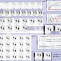
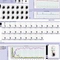
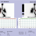
To avoid unnecessary radiation and for the less difficulties of the method, some investigators prefer ultrasonography as the initial imaging procedure. But the use of ultrasonography without scintigraphy in the diagnosis of the different etiologies in primary CH could be incomplete. This image method cannot always detect lingual and sublingual thyroid ectopy and is highly observer-dependent.
Combining scintigraphy and thyroid ultrasound in CH patients helps to:
Improve diagnostic accuracy
Identify a eutopic gland which may be normal, enlarged, or hypoplastic, guiding other investigation exams such as molecular studies
Prevent the incorrect diagnosis of athyreosis in the presence of no uptake on scintigraphy when ultrasound shows normal gland in situ
Detect thyroid ectopy reliably
22.3 Hyperparathyroidism
Hyperparathyroidism exists in three different forms: primary, secondary, and tertiary.
Diagnosis of primary hyperparathyroidism (PHPT) is characterized by suggestive clinical presentation and laboratory investigations (hypercalcemia and elevated serum concentrations of intact PTH). PHPT is a rare disease in adolescents, and limited data exist on pediatric and adolescent patients with primary hyperparathyroidism.
The causes of PHPT in the adolescent population include parathyroid adenomas, multiglandular disease (MGD), and parathyroid carcinoma. Neonatal hyperparathyroidism is a rare disorder, and it has been classified as a distinct disease entity; it is caused by inactivating calcium-sensing receptor (CASR) mutations that result in life-threatening hypercalcemia and metabolic bone disease.
Stay updated, free articles. Join our Telegram channel

Full access? Get Clinical Tree


