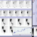
Fig. 15.1
(a, b) Scintigraphic study is performed after oral liquid meal administration revealing a progressive gastric emptying without gastroesophageal reflux. Time/activity curves corresponding to stomach and esophagus allow to calculate quantitative parameters (showing normal gastric emptying) and to confirm absence of gastroesophageal reflux, respectively. (c) The qualitative evaluation of dynamic study shows a normal oropharyngoesophageal transit after a single and complete swallowing. This finding is confirmed by time/activity curves analysis showing normal pattern of each curve and the sequential coordinated peaks of the T/A curves derived from upper, middle, and lower esophagus. Complete clearance of activity from the esophagus at the end of dynamic sequence is evident with a normal transit time (inferior than 5–6 s from upper esophagus tract to stomach)
15.2.1.2 Case 15.2 Normal Gastric Emptying Associated with a Mild Single Gastroesophageal Reflux Episode
A 3-year-old female patient with neurological impairment, congenital hydrocephalus, recurrent nocturnal cough, food refusal, and growth failure.
Due to a clinical suspect of gastroesophageal reflux disease (GERD), the patient underwent upper gastrointestinal series that revealed gastroesophageal reflux, delayed gastric emptying, and hiatal hernia.
Conversely, Tc99m gastric scintigraphy showed a single episode of gastroesophageal reflux and normal gastric emptying (46 % in 60 min, T/2 = 66 min).
Upper endoscopy and 24-h esophageal pH monitoring were negative for GERD.
Therefore, the patient stopped proton pump inhibitors.
In this case, the complete assessment for GERD has ensured a correct diagnosis and has modified the medical treatment (Fig. 15.2).
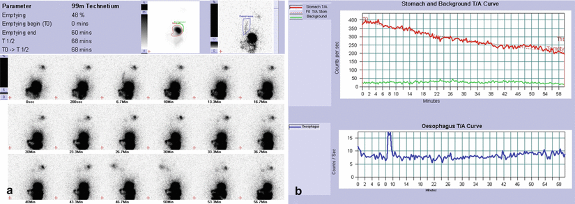
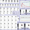
Fig. 15.2
(a, b) Gastric emptying study is performed after oral milk administration, and first dynamic study image allows to assess possible evidence of ab-ingestis inhalation (especially in children with neurological impairment and oral nutrition). The qualitative analysis of dynamic study reveals a progressive gastric emptying with a single gastroesophageal reflux episode that reaches upper esophageal tract. No evidence of abnormal activity is detectable corresponding to lung fields in the first frame and during dynamic registration. Time/activity curves corresponding to stomach and esophagus show normal gastric emptying and confirm a single mild gastroesophageal reflux episode with rapid washout
15.2.1.3 Case 15.3 Mild Delayed Gastric Emptying Associated with Severe Gastroesophageal Reflux Disease
A 4-year-old boy, born at term, cesarean section for polyhydramnios, postnatal diagnosis of esophageal atresia type C. The patient underwent surgical repair on the second day of life. The postoperative course was uneventful, and progressive oral alimentation was well tolerated. Treatment with H2 blocker was started and continued after discharge. After 2 months, anastomotic esophageal stenosis occurred, requiring endoscopic dilation. At the age of 4 months, H2 blocker was substituted by proton pump inhibitor (PPI). At the age of 14 months, the patient was evaluated for recurrent vomiting and cough. Upper GI endoscopy was unremarkable (patent esophageal anastomosis and normal gastric and esophageal mucosa), and esophageal pH monitoring was suggestive for moderate gastroesophageal reflux. PPI treatment cycles were beneficial. One year later, upper GI endoscopy revealed hiatal hernia, with positive histology for mild esophagitis, and still abnormal esophageal pH monitoring. Subsequent clinical course was good on PPI continuous treatment. However, at the age of 4 years, upper GI endoscopy showed macroscopic distal esophagitis and confirmed hiatal hernia. Gastric emptying scintigraphy showed slightly delayed clearance of the labeled food, in association to frequent episodes of gastroesophageal reflux. Therefore, the patient underwent laparoscopic antireflux surgery (Nissen fundoplication) associated to pyloromyotomy, with uneventful postoperative course and follow-up (Fig. 15.3).
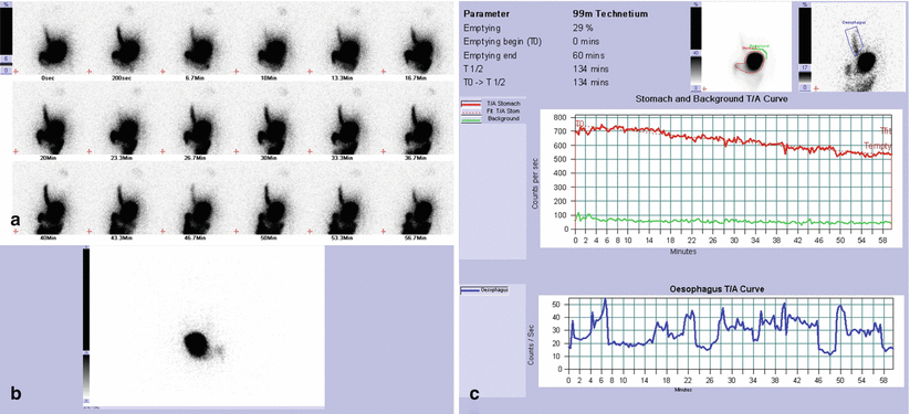
Fig. 15.3
(a–c) Qualitative analysis of dynamic study reveals a progressive gastric emptying with severe and frequent gastroesophageal reflux. Time/activity curves corresponding to stomach and esophagus show mild delayed gastric emptying (quantitative parameters: 29 % in 60 min, T/2 = 134 min) confirming evidence of numerous gastroesophageal reflux episodes. When gastric/esophagus activity ratio is higher than 3 %, gastroesophageal reflux is considered of large quantity. Number and volume of reflux help to define the grade of gastroesophageal reflux disease: in this clinical case, gastroesophageal reflux is considered as severe. Frame-by-frame assessment of dynamic study is necessary to evaluate reflux clearance (numerous gastroesophageal reflux episodes can be incorrectly detected by A/T curves and qualitative imaging as prolonged and slow reflux washout). (b) First study frame evaluation is important to highlight residual esophageal stasis after oral meal administration (in particular, in case of esophageal atresia history or surgical gastroesophageal treatment) differentiating from early reflux episode. It is always necessary for a careful assessment of possible evidence of activity corresponding to lung fields to exclude (or detect) ab-ingestis inhalation episodes
15.2.1.4 Case 15.4 Delayed Gastric Emptying Associated with Mild Gastroesophageal Reflux Disease (Scintigraphic Pretreatment Assessment)
A 3-year-old female, preterm (GE 24° sett, weight: 725 g; spontaneous delivery), polyhydramnios. Periventricular leukomalacia, bronchial dysplasia, retinopathy (laser therapy). Mild malnutrition, food refusal, dysphagia, chronic vomiting, and regurgitation.
Upper gastrointestinal (UGI) series: no UGI alterations, no gastroesophageal reflux (GER), delayed gastric emptying.
US pylorus: no alterations.
Tc99m gastric scintigraphy (prepyloric dilation): 17 % in 60 min, T/2 = 3.9 h, mild GER
Pyloric dilation: 12 mm hydrostatic pyloric dilation throughout endoscopy.
Clinical improvement and scintigraphic control after dilation were indicative of complete therapeutic efficacy, confirming diagnostic role of scintigraphy in pretreatment evaluation and monitoring therapeutic effect of pyloric dilation (Fig. 15.4).
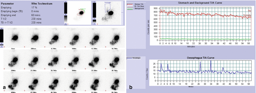
Fig. 15.4
(a) The qualitative analysis of dynamic study reveals a progressive but delayed gastric emptying with some associated gastroesophageal reflux episodes (almost five). Time/activity curves corresponding to stomach and esophagus show delayed gastric emptying (quantitative gastric parameters: 17 % in 60 min, theoretical T/2 = 3.9 h), confirming evidence of five mild gastroesophageal reflux episodes, respectively
15.2.1.5 Case 15.5 Delayed Gastric Emptying Associated with Severe Gastroesophageal Reflux Disease (Presurgical Assessment in Child Who Underwent Nissen Fundoplication and Pyloromyotomy)
An 8-year-old male patient with history of chronic iron-deficiency anemia (Hb 7.5 g/dl) and heartburn.
Upper endoscopy documented severe erosive esophagitis and hiatal hernia, and pH-multichannel intraluminal impedance (pH-MII) confirmed the diagnosis of severe gastroesophageal reflux disease (GERD). Treatment with proton pump inhibitors (PPI) was administered for 6 months with good clinical response and improvement of anemia.
An endoscopic re-evaluation without PPI documented esophagitis in the lower third of the esophagus.
Because of the dependence on medical therapy, surgical treatment was carried out.
In the preoperative period, patient underwent scintigraphy to evaluate gastric emptying and define the surgical procedure (Nissen fundoplication ± pyloromyotomy, (Fig. 15.5a, b).

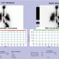
Fig. 15.5
(a, b) Qualitative analysis of dynamic study reveals a progressive but delayed gastric emptying with labeled meal stasis corresponding to stomach, up to the end of dynamic study. Numerous and severe gastroesophageal reflux episodes are also detectable without evidence of activity corresponding to lung fields. Time/activity curve corresponding to stomach shows delayed and irregular gastric emptying: after 32 min of latency time, gastric parameters are slower than normal (14 % in 60 min and theoretical T/2 = 2.4 h). Frame-by-frame and T/A esophagus curve analyses reveal numerous gastroesophageal reflux episodes: some of these show prolonged clearance, other ones rapid washout. (c, d) Laparoscopic Nissen fundoplication and pyloromyotomy
Tc99m gastric scintigraphy detected delayed gastric emptying (14 % in 60 min; T/= 2.4 h) associated to GER.
Laparoscopic Nissen fundoplication and pyloromyotomy were performed to treat GERD and improve gastric emptying. Currently, the patient is in good clinical condition with normal value of hemoglobin (Hb 12.5 g/dl).
15.2.1.6 Case 15.6 Delayed Gastric Emptying Associated with Severe Gastroesophageal Reflux Disease (In Child Who Underwent Nissen Fundoplication Without Scintigraphic Gastric Motility Assessment in the Preoperative Planning)
A 5-year-old male patient with history of severe gastroesophageal reflux disease (GERD) underwent Nissen fundoplication with placing of percutaneous endoscopic gastrostomy (PEG). A previous scintigraphic gastric emptying assessment was not performed. Persisting GERD symptoms, the child was referred to our center, and a gastric emptying scintigraphy was performed in the preoperative planning to choose adequate medical or surgical treatment. Delayed gastric emptying detected by gastric scintigraphy was considered the principal cause of failed previous Nissen fundoplication; a subsequent antireflux surgery associated with pyloromyotomy was necessary (Fig. 15.6).
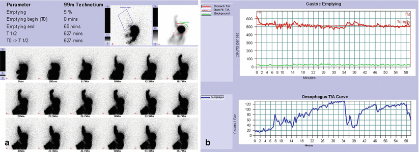
Fig. 15.6


(a, b) Gastric emptying study is performed after liquid meal administration by PEG tube. The qualitative analysis of dynamic study reveals poor gastric emptying with considerable retention of labeled meal in the stomach up to the end of dynamic study. Three severe gastroesophageal reflux episodes are also detectable without evidence of complete washout up to the end of dynamic registration. Time/activity curve corresponding to stomach shows severely delayed and irregular gastric emptying (5 % in 60 min with nonassessable T/2). Frame-by-frame and T/A esophagus curve analyses confirm evidence of three severe gastroesophageal reflux episodes with slow and partial washout
Related posts:


Stay updated, free articles. Join our Telegram channel
Full access? Get Clinical Tree
