Etiology
Incidence (%)
US findings
Diagnostic exams
Transient hydronephrosis
50–70
Isolated hydronephrosis (mild)
US
UPJO
10–30
Isolated hydronephrosis (DAP >15 mm)
MAG3
VUR
10–40
Intermittent hydronephrosis, ureteric dilatation, small kidney
VCUG/DMSA
UVJU
5–10
Ureteric dilatation
VCUG/MAG3
MCDK
1–5
Random renal cyst, little or no hyperechoic parenchima
DMSA
PUV
2–5
Thickened bladder wall, hydronephrosis, hyperechoic kidney, ureteric dilatation, oligoidramnios (fetal), key-hole sign (fetal)
VCUG/MAG3
Other postnatal obstructive uropathies than PUJO can be associated with prenatal or neonatal hydronephrosis, such as posterior urethral valves (PUV) or ureterovescical junction obstruction (UVJO), and are more often associated with higher grades of hydronephrosis. Vesicoureteric reflux (VUR) is present in 5–15 % of prenatal hydronephrosis and, differently from the aforementioned obstructive uropathies, does not appear to be related to the severity of the dilatation, which is obvious if one thinks to the intermittent nature of VUR. As a consequence, postnatal imaging could be warranted even in the setting of mild prenatal hydronephrosis.
4.1.2 Postnatal Management
There is general consensus that moderate to severe prenatal hydronephrosis should undergo routine postnatal imaging: in such cases, the risk of postnatal pathological finding ranges from 45 to 80 % of cases (the more severe hydronephrosis is, the higher is the risk). In infants with mild hydronephrosis, the role of postnatal imaging is less clear; however, up to 10 % of these cases may have significant uropathies. Therefore, even in infants with mild or disappeared hydronephrosis, at least a single postnatal imaging seems of benefit.
Renal ultrasonography (US) is the principal imaging technique used in both postnatal and in prenatal life, due to the easy availability, low cost, and absence of radiation exposure; however, it is operator-dependent.
Postnatal ultrasonography should be performed at 3–7 days of life, in order to avoid underestimation or incorrect assessment due to physiological disidratation occurring in the very first days of life.
Differential diagnosis of postnatal hydronephrosis based on US findings is shown in Table 4.1.
When dilatation of calyces and pelvis is isolated and not associated with any other abnormal finding, up to 50–70 % of these cases will be transient, and will gradually disappear spontaneously without any clinical sequelae or renal impairment. The remaining 30 % of prenatal hydronephrosis will require further postnatal investigation other than ultrasonography.
4.1.3 Diagnostic Investigations
Since the vast majority of hydronephrosis will gradually disappear, periodic urinary tract ultrasonography (every 2–3 months in the first year of life) is mandatory in order to assess the evolution of the urinary dilatation.
In cases of hydronephrosis associated with ureteric dilatation, and in patients presenting with symptomatic UTI, international guidelines recommend the execution of VCUG in order to rule out the presence of VUR, as in the chapter on VUR. Otherwise, in asymptomatic isolated hydronephrosis, routine VCUG may not be needed: in this instance, infact VUR prevalence is low (15 %), with VUR being of low grade in most cases and clinically not significant.
In the absence of VUR, and when periodic ultrasonography shows persistence or worsening of hydronephrosis, the presence of UPJO has to be suspected. Dynamic renal scan is the imaging investigation of choice.
The most popular dynamic scan in cases of suspected obstruction uses Tc-mercaptoacetyltriglycine (MAG3) as radiotracer. Briefly, MAG3 is primarily excreted by secretion from the proximal tubule. As a result, it can allow evaluation of both split renal function and urine progression to the bladder.
Renal immaturity prompts us to perform nuclear medicine exams before the infant is 1 month old, for the risk otherwise to underestimate split renal function. For the same reason, the child must be adequately fed and well hydrated. IV fluids are however considered unnecessary to the vast majority of cases. Sedation is not required, and the placement of a bladder catheter should be reserved to much selected cases.
4.1.4 Particular Cases
4.1.4.1 Symptomatic Hydronephrosis
Symptomatic hydronephrosis is a totally different entity from congenital asymptomatic hydronephrosis. It generally occurs in a school-age child or adolescent, which presents with intermittent abdominal pain, sometimes together with vomiting. Urinary tract infection may or may not be present. Perinatal US may be negative, or a previous hydronephrosis may have solved spontaneously. Children as young as 3 years may present with colicky pain due to hydronephrosis. In the vast majority of symptomatic cases, aberrant vessels may be found going to the lower pole of the kidney and causing intermittent compression of the UPJ. In situation of hyperdiuresis, the UPJ is compressed and the pelvis gets distended; this in turn determines reduction of diuresis and reduction of the hydronephrosis. The pain is therefore intermittent, as is the US finding of hydronephrosis, Nonetheless, this mechanism can lead to significant loss of kidney function. Grade of hydronephrosis can be extremely variable at US. MAG3 scan can be nonobstructive. The treatment is straightforward surgical.
4.1.4.2 Hydronephrosis in Renal Anomalies
Three peculiar cases are possible: hydronephrosis in duplex system, hydronephrosis in ectopic kidney, and in horseshoe kidney. In most cases of renal anomalies, an anatomical defect of UPJ is present and spontaneous resolution of obstruction is less plausible.
Hydronephrosis in horseshoe kidney occurs more commonly than in normal kidneys. Vascular anomalies with aberrant vessels may be present as well as the anomalies of the UPJ with very high insertion of the ureter on the pelvis. The surgical approach must be decided, keeping in mind the abnormal position of the renal pelvis (anterior and medial to the kidney), and a laparoscopic transperitoneal approach should be considered nowadays the surgical approach of choice. Section of the hystums is no longer an option.
Hydronephrosis in the ectopic kidney can be associated with reduced function due to renal hypoplasia. At US, the pelvis is generally malrotated. Evaluation of UPJO and indication to surgery may not be so evident.
Hydronephrosis in the duplex kidney is the less common. It generally occurs in the lower pole. Compression by aberrant vessels to the lower pole can be a possible cause. Hypoplasia of UPJ in case of incomplete duplex system (two pelvis draining in a single ureter) is also a possible occurrence.
4.2 The Surgical Treatment
Surgical correction of UPJO consists of excision of the obstructed tract at the UPJ and a pyeloureteral anastomosis, as described by Anderson–Hynes technique. Surgery can be performed in an open fashion, through a lumbar lateral, anterior or posterior incision. In the last decade, minimally invasive approaches have become popular, which means by small open-fashioned incisions in infants and young children, laparoscopy-assisted procedures in infants and young children, laparoscopic trans-or retroperitoneal in grown-up children, or robotics-assisted procedures. These procedures have gained wide acceptance due to the reduced pain, quicker recovery, and similar results as in traditional open surgery.
In children and adolescents with aberrant vessels, cephalad mobilization of the vessel (so-called “vascular hitch”), according to Hellstrom technique, can be as effective as the dismembered Anderson–Hynes pyeloplasty in selected patients.
4.3 The Follow-Up
Periodical US is recommended after surgery. Significant reduction of the hydronephrosis is expected, but it may take months or years to take place. Persistent dilatation of the renal pelvis is a possible occurrence even in the absence of obstruction, especially when very large hypotonic pelvis and calyces were observed before surgery. MAG3 scan should be repeated at 6–12 months follow-up, or earlier if dilatation persists. In this latter case, definition of persistent obstruction from large hypotonic pelvis with dilated calyces can be particularly challenging for the nuclear medicine specialist.
4.4 Dynamic Renal Scintigraphy
Dynamic renal scintigraphy allows to assessing renal function and drainage, evaluating both renal excretion and differential renal function (DRF).
Most common indications in pediatric population include determination of DRF and drainage in hydronephrosis and megaureter, in diagnostic phase, after surgical repair (when performed), and during follow-up.
4.4.1 Study Technique and Interpretation
The patient, adequately hydrated, receives 99mTc-MAG3 (mercaptoacetyltriglycine) as a bolus injection contextually with the starting of 30-min dynamic acquisition The administered activity of radiotracer is adjusted to the patient’s weight, according to EANM dosage card and to the national regulations.
Region-of-interest (ROI) around the heart, the kidneys, and the area around the kidneys (“background”) are drawn on a summed image of dynamic frames using a dedicated software; thus, time/activity curves are obtained, of expression of perfusion (flow T/A) and of renal function and drainage (renograms), respectively. In renograms, the ascending part represents the parenchymal phase (single kidney function) and the descending part reflects renal transit and emptying; with the same method, differential renal function (DRF or split function) is also determined.
If renal drainage is impaired at the end of the dynamic study, an additional image after 5 min standing upright is acquired (“gravity-assisted drainage,” GAD1). In case of still poor drainage (cutoff = 30 %), a 1 mg/kg dose of furosemide is given intravenously and an additional 20-min dynamic acquisition follows. If pyelic drainage is still incomplete (half-time >20 min, drainage <30 %), a second “gravity-assisted drainage” test (GAD-2) is also performed. The drainage is classified as impaired on the basis of all the previous mentioned three tests.
4.4.2 Teaching Cases
4.4.2.1 Case 4.1 Normal Ultrasonographic and Scintigraphic Pattern After Left Pyeloplasty
A 5-year-old boy came to Emergency for recurrent left lumbar pain. On ultrasonography, grade 3 left hydronephrosis was detected. No urinary tract dilatation was referred at prenatal ultrasound. Dynamic renal scintigraphy showed symmetrical split function with obstructive urinary drainage at diuretic test. Pyeloplasty for correction of UPJO was performed with complete ultrasonographic resolution of the left hydronephrosis. MAG3 scan was repeated at 1-year follow-up after pyeloplasty (Fig. 4.1).
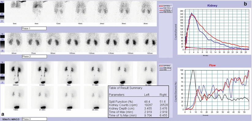
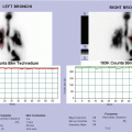
Fig. 4.1
MAG3 dynamic renal scan (after pyeloplasty): dynamic images (a) show good and homogeneous radiotracer uptake and normal drainage in both kidneys; renograms (b) confirm normal function and drainage of both kidneys (left DRF: 48 %; right DRF: 52 %); flow T/A curves (b) show synchronous and symmetrical perfusion
4.4.2.2 Case 4.2 Persistent nonobstructive Left Hydronephrosis with Normal Split Function
A boy of 3 years was followed for prenatal diagnosis of bilateral hydronephrosis, stable during pregnancy and confirmed at birth with an anteroposterior diameter (APD) of 15 mm. After birth, progressive decrease of right pyelic dilatation and persistent left hydronephrosis was found. One episode of acute pyelonephritis occurred at 3 months with subsequent cystography (VCUG) negative for vesicoureteric reflux. No further episodes of urinary tract infections (UTI). At the age of 3 years, he presented persistent left hydronephrosis with anteroposterior (AP) pyelic diameter of 24 mm and good parenchymal thickness, asymptomatic. MAG3 scan showed normal left split renal function and adequate urinary drainage after GAD-1 test. No surgical indication. Ultrasonographic follow-up was recommended (Fig. 4.2).
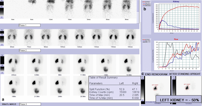
Fig. 4.2


MAG3 dynamic renal scan: dynamic images (a) show good radiotracer uptake and nonhomogeneous intraparenchymal distribution in left kidney; an area devoid of tracer corresponding to dilated renal pelvis is evident and drainage is poor; right kidney presents good and homogeneous radiotracer uptake and normal drainage; renograms (b) confirm normal function but poor drainage of left kidney (“plateau pattern”) and normal function and drainage of right kidney (left DRF: 53 %; right DRF: 47 %); flow T/A curves show synchronous and symmetrical perfusion. Gravity-assisted drainage-1 test shows significant emptying of left kidney, excluding a significant obstacle in the upper pyelic junction (c)
Related posts:


Stay updated, free articles. Join our Telegram channel
Full access? Get Clinical Tree
