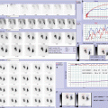
Fig. 5.1
MAG3 dynamic renal scan: dynamic images (a) show good and homogeneous radiotracer uptake and normal drainage in both kidneys, with mild and transient radioactivity in both ureters; renograms (b) confirm normal function and drainage of both kidneys; flow T/A curves (b) show synchronous and symmetrical perfusion. Ureter T/A curves (c) show normal pattern
5.1.2.2 Case 5.2: Nonrefluxing and Nonobstructive Primary Right Megaureter with Normal Split Renal Function
Female patient affected by prenatally detected right uropathy.
At 1 month of life, the ultrasonographic scan showed a right urinary tract dilation (pelvic anteroposterior diameter of 6 mm; distal ureteral dilation of 5 mm).
At the follow-up, the patient had a febrile urinary infection and a worsening of the right ureteral dilation (up to 10 mm); for this reason, the voiding cystourethrography was performed, but it revealed the absence of urinary pathologies.
The following technetium-99m-labeled mercaptoacetyl-triglycine (MAG-3) renography detected a normal split renal function with evidence, at the right side, of the megaureter and slow urinary drainage but absence of obstructive urinary pattern (no diuretic injection was necessary).
At the last clinical checkup, the patient remained asymptomatic and the ultrasonographic scan showed the stability of the right megaureter (diameter of 10 mm).
The management of the patient is actually based on wait-and-see strategy associated to serial ultrasonographic evaluations (Fig. 5.2).

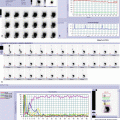
Fig. 5.2
MAG3 dynamic renal scan: dynamic images (a) show good and homogeneous radiotracer uptake and normal drainage of left kidney; right kidney presents good and homogeneous radiotracer uptake; pyelic drainage is poor, and persistent radioactivity stasis is evident also in ipsilateral dilated ureter; renograms confirm normal function and drainage of left kidney, and normal function, prolonged transit time, and delayed excretion of right kidney; flow T/A curves (b) show synchronous and symmetrical perfusion. Gravity-assisted drainage-1 test shows significant emptying of right kidney and right ureter (c). This finding is confirmed by ureter T/A curves (d): normal pattern of left ureter and dilated nonobstructive pattern of right ureter
5.1.2.3 Case 5.3: Nonobstructive Right Megaureter with Low Right Split Renal Function, Autosomal Dominant Bilateral Polycystic Kidney Disease, and Bilateral Urinary Microlithiasis
A 15-year-old girl with history of acute renal pain episodes, bilateral urinary microlithiasis, and poor right renal function was admitted to our hospital.
The initial ultrasonographic scan showed right kidney hypoplasia, bilateral urinary microlithiasis, bilateral polycystic kidney disease, and right urinary tract dilation.
The following technetium-99m-labeled mercaptoacetyltriglycine (MAG-3) renography revealed a poor right split renal function and right kidney hypoplasia with evidence at the right side of megaureter and slow urinary drainage but absence of obstructive urinary pattern (no diuretic injection was necessary).
At the last clinical follow-up, the patient remained asymptomatic.
The management of the patient is actually based on clinical and ultrasonographic evaluations (Fig. 5.3).

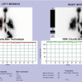
Fig. 5.3
MAG3 dynamic renal scan: dynamic images (a) show good and homogeneous radiotracer uptake and normal drainage of left kidney; right kidney is smaller than the other one, with irregular shape; right kidney presents reduced radiotracer uptake with nonhomogeneous intraparenchymal distribution due to an area devoid of tracer corresponding to dilated collecting system, and the drainage is poor; persistent radioactivity is also evident in ipsilateral ureter. Renograms (b) show normal function and drainage of left kidney and severe hypofunction, prolonged transit time, and delayed excretion phase of right kidney; flow T/A curves (b) show synchronous but asymmetrical perfusion (reduced in right kidney). Gravity-assisted drainage-1 test shows significant emptying of right kidney and right ureter (c). This finding is confirmed by ureter T/A curves (d): normal pattern of left ureter and dilated nonobstructive pattern of right ureter
5.1.2.4 Case 5.4: Secondary Left Megaureter by Posterior Urethral Valves
Male infant affected by prenatally detected left uropathy.
During the first month of life, the initial ultrasonographic scan showed a left urinary tract dilation (pelvic anteroposterior diameter of 7.5 mm; distal ureteral dilation of 16 mm) and a bladder wall hypertrophy.
The voiding cystourethrography was performed, and it revealed the absence of vesicoureteral reflux but the presence of posterior urethral valves; for this reason, at 6 months of life, the patient was submitted to endoscopic ablation of posterior urethral valves.
The ultrasonographic scan at 2 months postoperative follow-up confirmed the stability of the preoperative left megaureter.
The following technetium-99m-labeled mercaptoacetyl- triglycine (MAG-3) renography showed a normal split renal function with left ureteral dilation and poor urinary drainage that required the diuretic injection; no obstructive urinary pattern was detected after the diuretic injection.
At the last day hospital follow-up, the patient remained asymptomatic, and the ultrasonographic scan revealed the reduction of the left megaureter (diameter of 8 mm).
Conservative management is actually the utilized strategy for the patient (Fig. 5.4).

Fig. 5.4


MAG3 dynamic renal scan: dynamic images (a) show good radiotracer uptake in left kidney with nonhomogeneous intraparenchymal distribution due to an area devoid of tracer corresponding to dilated renal pelvis and collecting system; drainage is poor, with persistent radioactivity also in left ureter, in particular in its distal tract; right kidney presents good and homogeneous radiotracer uptake and normal drainage; renograms (b) confirm normal function but mildly delayed excretion phase of left kidney, and normal function and drainage of right kidney; flow T/A curves (b) show synchronous and symmetrical perfusion. Gravity-assisted drainage-1 test shows no significant washout of left kidney and left ureter (c). Diuretic dynamic images (d) and diuretic renogram (e) show prompt and significant washout in both left kidney and left ureter after administration of furosemide. Ureter T/A curves (f) show: normal pattern of right ureter and dilated nonobstructive pattern of left ureter during dynamic renal scan (upper graph); normal pattern of right ureter and prompt washout of left ureter during diuretic scintigraphy (lower graph)
Related posts:


Stay updated, free articles. Join our Telegram channel
Full access? Get Clinical Tree
