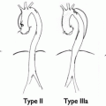
Fig. 23.1
Type II endoleak
(b)
Type II-b—perfusion from lumbar arteries
3.
Type III—perfusion resulting from a defect within the graft:
(a)
Type III-a—between the components of a modular graft
(b)
Type III-b—tear or other disruption within graft fabric
4.
Type IV—generalized, diffuse leakage due to fabric porosity
5.
Type V—persistent pressurization or enlargement of the aneurysm sac without demonstrable flow or endoleak. Because no flow can be visualized (by definition), the best term for this situation is “endotension.”
5 Diagnosis
As an endoleak creates continued pressurization of the aneurysm sac, the potential for rupture still exists. Because the chance of this varies according to the type and timing of the leak (as leaks can occur late), routine follow-up with imaging of the aneurysm repair remains the standard of care.
5.1 Clinical Follow-up
No standardized algorithm exists. Most often, patients undergo initial postoperative imaging and clinical evaluation approximately 1 month after their surgery. Subsequent imaging and evaluation is performed are 6 and 12 months, and if all is well, annually thereafter. If an abnormality such as a newly diagnosed endoleak, persistent endoleak, or aneurysm sac enlargement is detected during routine surveillance, further evaluation is necessary.
5.2 Imaging Modalities
The goal of postoperative imaging is to assess the size of the residual sac and to identify or exclude the presence of an endoleak. Each can be accomplished by computerized tomography (CT), magnetic resonance (MR) scanning, or ultrasound (USG). Angiography is frequently used to look for (and treat when applicable) the source of an endoleak. However, it cannot assess the sac size and is obviously much more invasive; therefore, it is not used for routine postoperative surveillance.
5.2.1 Computerized Tomography
Often combined with postprocessed vessel reconstruction (CT angiography CTA), this is the primary study used for postoperative surveillance because of its high resolution. CTA is felt by most to be the most sensitive test for detecting endoleaks when compared to both USG and conventional angiography. Other advantages include safety, availability, reproducibility, accuracy, lack of operator dependence, ease of interpretation, and rapid acquisition times. Importantly, however, significant disadvantages exist. CTA requires that the patient be exposed to a not-insignificant dose of ionizing radiation on an annual basis for life. Further, it requires a relatively high dose of intravenous contrast dye with risks of allergic reaction and nephrotoxicity; the latter effect having been shown to be associated with cumulative renal damage over time. Other disadvantages include difficulty in discerning calcifications from contrast and the directionality of blood flow from an endoleak. Further, subtle late leaks are poorly visualized. Finally, the cost of a CT scan is greater than other available imaging modalities. Timing the scan (i.e., precontrast, and postcontrast) can help to identify fixed calcium and late endoleaks; however, the radiation dose needed is obviously greater. Overall, CT and CTA provide the highest resolution but have significant drawbacks to their use.
5.2.2 Magnetic Resonance Angiography
Recent data suggest that magnetic resonance angiography (MRA) is as sensitive at detecting endoleaks as CT. While appealing due to the elimination of nephrotoxic contrast agents (in most situations) and radiation, the disadvantages of MRA are significant enough to preclude its routine use for postoperative surveillance. First, grafts made of stainless steel (Cook Zenith, Bloomington, IN) are in theory not compatible, although empirically no adverse events have been seen in such patients undergoing early or late MR scanning. Further, many patients are unable to undergo MR scanning due to preexisting implants. Most clinicians feel that MR does not image the central vascular system as well as other imaging modalities. Finally, MR suffers from the well-known drawbacks of longer acquisition time and patient dissatisfaction (noise and claustrophobia), further limiting its role for widespread routine surveillance in this situation.
5.2.3 Ultrasonography
Although reports vary extensively regarding the sensitivity in which ultrasonography (USG) can detect endoleaks (43–100%), it has many advantages that make it an ideal screening tool. Foremost, it is a safe, noninvasive, inexpensive tool that is highly reproducible given the proper environment. No contrast is needed and there is no exposure to radiation. However, the examination does take longer to complete and is subject to a greater degree of operator/interpreter variability. Also, patient variables such as body habitus, fasting status, and bowel gas can make the exam less accurate. Patients therefore should be instructed to fast before the exam, and lower frequency (3–5 Hz) scan heads should be utilized to allow for deeper penetration. Finally, it should be obvious that if USG results are to be reliably trusted, they should be obtained by a very experienced, accredited laboratory with rigorous quality control and internal verification of results.
Some argue that USG is less sensitive than CT in detecting endoleaks. This may or may not be true, but it has been pointed out by many that if a sac is shrinking, the presence of a Type II endoleak per se is not critical. Ultrasound is highly accurate for determining sac size, and thus an increasing number of clinicians and investigators are relying primarily upon USG for routine postoperative follow-up, resorting to CT (and/or angiography) only if a problem is identified by USG.
5.2.4 Conventional Angiography
Because of the risks and expense of arterial access, this is the most invasive imaging modality used in the detection of postoperative endoleaks. In addition, it too requires radiation and the administration of a contrast agent (although with selective catheterization, the operator has more control of the actual dosage used and can limit the amount when mitigating factors exist). It cannot determine the sac size and has been shown to be less sensitive in detecting endoleaks. For all of these reasons, it is not used as a postoperative surveillance tool. However, if an endoleak is detected, angiography is necessary if intervention is planned and as such should be viewed as more of a therapeutic rather than diagnostic tool.
Related posts:

Stay updated, free articles. Join our Telegram channel

Full access? Get Clinical Tree


