Endolymphatic sac tumor – immediate postoperative radiosurgery of surgical bed
SKULL BASE REGION
Cerebellopontine angle/petrous bone
HISTOPATHOLOGY
Endolymphatic sac tumor
PRIOR SURGICAL RESECTION
Yes
PERTINENT LABORATORY FINDINGS
N/A
Case description
The patient is a 74-year-old female with a known history of an endolymphatic sac tumor (ELST) involving the left cerebellopontine angle (CPA) and temporal bone leading to hearing loss, which was previously resected about 4 years earlier at another institution. Gross total resection (GTR) was achieved, but her perioperative course was complicated by a temporal stroke and aphasia, requiring long-term rehabilitation and speech therapy. She presented to our care with a new left facial weakness. Follow-up brain magnetic resonance imaging (MRI) showed local tumor recurrence in the left CPA, with extension to the petrosal bone ( Figure 7.33.1 ). Repeat surgical resection of the recurrent tumor was recommended, and the patient agreed to proceed. Immediately prior to the surgery, her facial weakness had already progressed to a complete facial palsy. Due to tumor invasion and complete facial palsy, a complete mastoidectomy/petrosectomy was performed. The specimen confirmed ELST recurrence without any new histopathological changes, and postoperative MRI confirmed GTR ( Figure 7.33.2 ). Postoperatively, the patient remained neurologically stable, with left complete facial palsy and hearing loss. Given tumor recurrence despite initial GTR, stereotactic radiosurgery (SRS) of the surgical bed was recommended immediately after the second surgery. The patient received 20 Gy in 5 fractions on the TomoTherapy unit. ( Figure 7.33.3 ).
Radiosurgery Machine
TomoTherapy
Radiosurgery Dose (Gy)
20, at the 95% isodose line
Number of Fractions
5
Figure 7.33.1.
Coronal and axial postcontrast T1-weighted images revealing a left-sided contrast enhancing lesion in the cerebellopontine angle with invasion of the petrous bone, which corresponds to recurrence of the previously resected endolymphatic sac tumor.
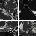
Figure 7.33.2.
Postcontrast coronal T1-weighted fat saturated, and postcontrast coronal and axial T1-weighted images performed after second surgery via a left translabyrinthine approach for tumor recurrence, confirming gross total resection. The fat packing in the resection cavity is clearly noted without any surrounding contrast enhancement.
Only gold members can continue reading. Log In or Register to continue


 Esthesioneuroblastoma – delayed postoperative radiosurgery for recurrence at long-term
 Null cell – delayed postoperative radiosurgery for growing perioptic residual
 Chondrosarcoma – definitive radiosurgery after subtotal resections
 Trigeminal neuralgia due to microvascular conflict – upfront radiosurgery
 Capillary hemangioma – postoperative radiosurgery for residual tumor
 Superior sagittal sinus meningioma – delayed postoperative, multisession radiosurgery for growing residual


