Epidural Abscess


Clinical Presentation


The patient is a 51-year-old male with 3-week history of mid back pain with radiation initially to the right side, but more recently the pain has migrated to the left side. The pain is moderate to severe in intensity. He has become weak in the legs in the past 36 hours, and over the past 12 hours he has become unable to move his lower extremities. He has no bladder or bowel dysfunction. The patient has a sensory deficit (absent pin prick sensation) at and below the T6 level. He has had intermittent fever for the past 3 weeks. Blood cultures confirmed presence of methicillin-resistant Staphylococcus aureus (MRSA) septicemia.


Imaging Presentation


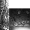
Magnetic resonance (MR) imaging revealed a fusiform right posterolateral epidural fluid collection with peripheral enhancement, extending from the T6 to T8 level ( Figs. 28-1 to 28-3 ) . The MR appearance is consistent with epidural abscess. The abscess is displacing the spinal cord anteriorly and to the left (see Fig. 28-2 ). A decompressive laminectomy was performed from T6 to T8 levels to débride and evacuate the thoracic epidural abscess. The patient was placed on intravenous antibiotics for 6 weeks, which was administered via a peripherally inserted central catheter (PICC) line.


Figure 28-1


Posterior Epidural Abscess, Mid-Thoracic Spine.

Sagittal fat saturated T2-weighted image. The abscess ( A ) is predominantly T2 hypointense centrally with surrounding fluid. The dura ( obliquely oriented arrows ) is displaced anteriorly by the posterior epidural abscess. The spinal cord ( C ) is displaced anteriorly and contains T2 hyperintense edema ( E ).


Figure 28-2


Posterior Epidural Abscess, Mid-Thoracic Spine.

Same patient as in Figure 28-1 . The epidural abscess ( arrows ) is predominantly T2 hyperintense on axial gradient echo image. The abscess displaces the spinal cord ( C ) anteriorly and to the left.


Figure 28-3


Posterior Epidural Abscess.

Same patient as in Figures 28-1 and 28-2 . T1-weighted sagittal MR images before (image A ) and after (image B ) IV contrast administration. The abscess is T1 isointense ( arrows in image A ) with respect to the spinal cord and displaces the cord anteriorly. In image B , the abscess ( A ) does not enhance centrally but enhances at its margin. There is also enhancement in the adjacent epidural space and dura ( arrows ).


Discussion


Spinal epidural abscess (epidural empyema) is an infection in the epidural space. This begins as a phlegmon that cavitates and fills with infectious liquid and necrotic debris. These abscesses most commonly arise in the anterior epidural space in the lower thoracic or lumbar region secondary to disc space infection and vertebral osteomyelitis (spondylodiscitis). Epidural abscess may also occur in the cervical or upper thoracic spinal canal secondary to spondylodiscitis or hematogenous dissemination ( Figs. 28-4 and 28-5 ) . Cervical epidural abscesses more commonly occur in the anterior epidural space, whereas thoracic epidural abscesses more commonly arise posteriorly (see Figs. 28-1 to 28-3 ).


Figure 28-4


C6-7 Discitis, Osteomyelitis, and Anterior Paraspinal Phlegmon.

On sagittal T1-weighted MR image A , the prevertebral phlegmon ( short arrows ) and anterior epidural phlegmon ( long arrows ) are relatively isointense compared to the spinal cord. On sagittal contrast-enhanced image B , the prevertebral phlegmon ( short arrows ) enhances diffusely. The enhancing anterior epidural phlegmon ( long arrows ) extends inferiorly into the upper thoracic anterior epidural space, displacing the spinal cord posteriorly.


Figure 28-5


C6-7 Discitis, Osteomyelitis, and Anterior Paraspinal Phlegmon.

Axial contrast enhanced image at C6-7 level. The phlegmon ( P ) in the anterior epidural space deforms the ventral aspect of the thecal sac and spinal cord. The anterior paraspinal phlegmonous tissue displays ill defined enhancement ( short arrows ).


Anterior epidural abscesses associated with spondylodiscitis are usually centered at an intervertebral disc level, but may extend over one to several vertebral levels. Posterior epidural abscesses occur by direct extension from paraspinal infections, hematogenous spread, or via the paravertebral venous plexus ( Figs. 28-6 and 28-7 ) . Posterior epidural abscesses may occur by direct extension after surgery, spinal anesthesia, or percutaneous spinal injections. Anterior or posterior epidural abscesses may arise from infections in the oral cavity, neck soft tissues, lungs, gastrointestinal (GI) tract (diverticulitis), genitourinary (GU) tract (bladder infection or pyelonephritis), from endocarditis, or secondary to septicemia. Spinal epidural infections may disseminate through the spinal canal diffusely along the leptomeninges.


Figure 28-6


L3-4 Discitis, Osteomyelitis, Paraspinal Phlegmon, and Posterior Epidural Abscess.

Infecting organism Streptococcus viridans . On unenhanced T2-weighted image A , the fluid and inflammation in the L3-4 disc is T2 hyperintense. The adjacent vertebral marrow edema and inflammation is T2 hyperintense ( short arrows in image A ). On contrast-enhanced fat-saturated T1-weighted MR image B , the vertebral marrow ( short arrows ) adjacent to the L3-4 disc enhances. Tissue within the L3-4 intervertebral disc ( long arrow in image B ) enhances with contrast. The posterior epidural abscess, which extends superiorly from the L3-4 level, is T2 hyperintense ( long arrows in image A ) and the abscess contains nonenhancing liquid spaces and enhances intensely at its margin ( long arrows in image B ).


Figure 28-7


L3-4 Discitis, Osteomyelitis, Paraspinal Phlegmon, and Posterior Epidural Abscess.

Same patient as in Figure 28-6 . Axial fat-saturated contrast-enhanced MR images A and B . The abscess ( arrow in image A ) is positioned posteriorly and along the right lateral margin of the thecal sac. The abscess contains nonenhancing liquid. In image B , at the L3-4 level, there is a right anterior and lateral enhancing phlegmonous tissue ( small arrows ) containing a small nonenhancing abscess ( A ).

Only gold members can continue reading. Log In or Register to continue

Stay updated, free articles. Join our Telegram channel

Aug 25, 2019 | Posted by in NEUROLOGICAL IMAGING | Comments Off on Epidural Abscess

Full access? Get Clinical Tree

Get Clinical Tree app for offline access