Esophageal cancer is an uncommon malignancy that ranks sixth in terms of mortality worldwide. Squamous cell carcinoma is the predominant histologic subtype worldwide whereas adenocarcinoma represents the majority of cases in North America, Australia, and Europe. Esophageal cancer is staged using the American Joint Committee on Cancer and the International Union for Cancer Control TNM system and has separate classifications for the clinical, pathologic, and postneoadjuvant pathologic stage groups. The determination of clinical TNM is based on complementary imaging modalities, including esophagogastroduodenoscopy/endoscopic ultrasound; endoscopic ultrasound–fine-needle aspiration; computed tomography of the chest, abdomen, and pelvis; and fluorodeoxyglucose PET/computed tomography.
Key points
- •
Squamous cell carcinoma and adenocarcinoma represent more than 90% of cases of esophageal cancer, the latter of which is the most prevalent histologic subtype in North America.
- •
The most commonly used scheme for staging esophageal cancer is the eighth edition of the American Joint Committee on Cancer/The International Union for Cancer Control TNM system.
- •
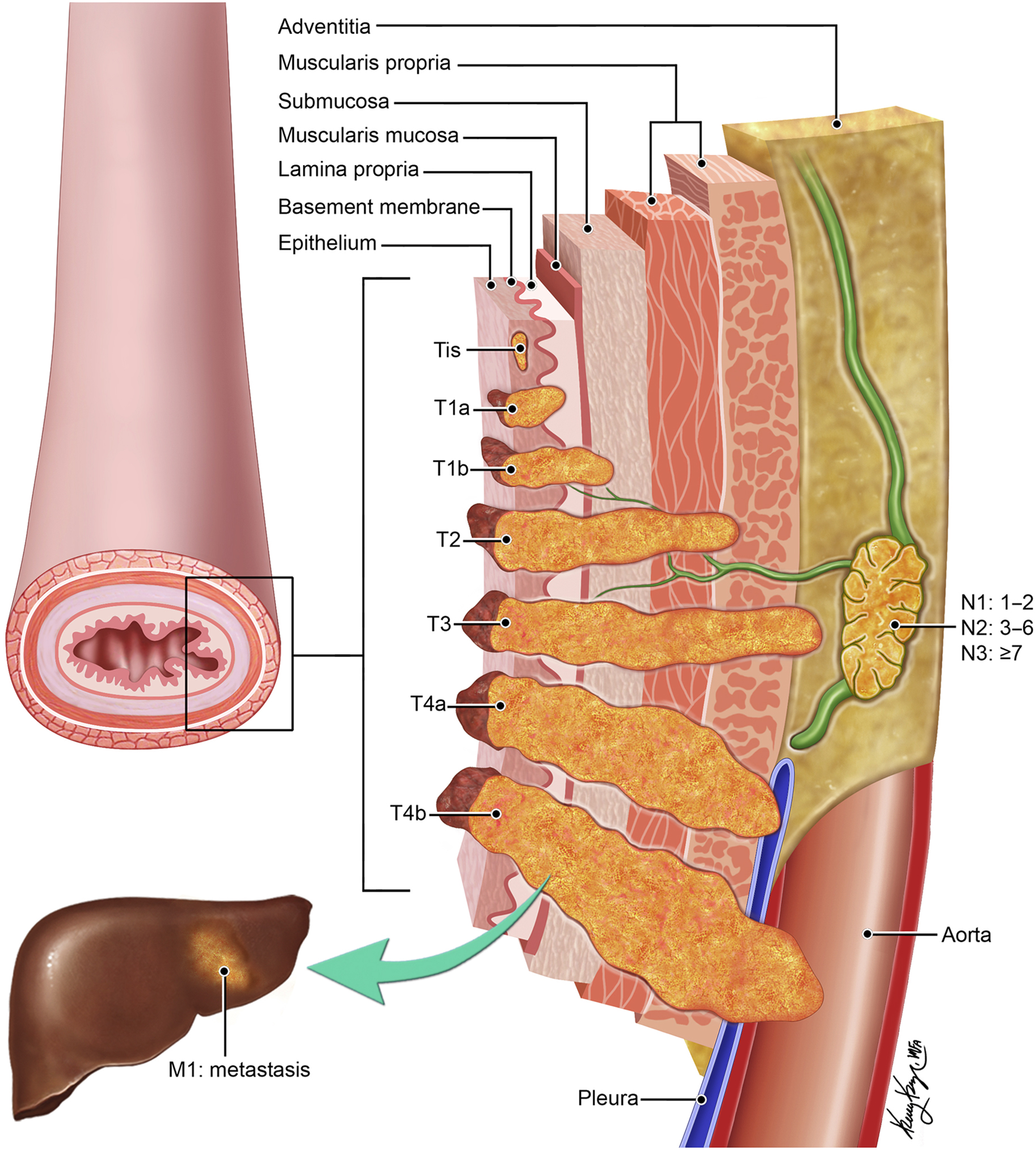
The T category ranges from Tis (high-grade dysplasia) to T4 (invasion of the primary tumor into adjacent structures).
- •
The N category is subdivided into the following components based on the number of involved regional lymph nodes: N1—1 to 2 lymph node metastases, N2—3 to 6 lymph node metastases, and N3—greater than 6 lymph node metastases.
- •
The M category includes M0 (no metastasis) and M1 (nonregional lymph nodal metastasis and distant visceral metastasis) subcategories.
Introduction
Esophageal cancer is a relatively uncommon malignancy in the United States, although its incidence has been increasing since the 1980s. It currently ranks seventh in terms of incidence and sixth in overall mortality worldwide. The 2 most common histologic types of esophageal cancer are squamous cell carcinoma (SCC) and adenocarcinoma (AC), representing more than 90% of all cases. SCC accounts for more than 80% of all cases worldwide and is the predominant histologic type in less developed countries. In contrast, AC represents more than 60% of all cases in North American, Australia, and Europe. ,
The treatment of esophageal cancer is stage-specific in order to ensure the best possible clinical outcomes. The treatment plan typically includes surgical resection for early disease, multimodality treatment with neoadjuvant chemotherapy, or combined chemoradiotherapy followed by surgery for patients with locally advanced cancer and systemic therapy for patients with metastatic disease. Accordingly, accurate pretreatment staging is important to ensure the development of appropriate treatment plans. The most commonly used staging for esophageal cancer is the eighth edition of the American Joint Committee on Cancer (AJCC)/The international Union for Cancer Control (UICC) TNM system. TNM staging includes determination of the depth of local invasion by the primary tumor (T), the presence and number of regional lymph nodes involved (N), and the presence or absence of distant metastasis (M). Because of differences in epidemiology, pathogenesis, location, and outcomes of the major histologic subtypes, TNM staging is separate for AC and SCC and takes into account the differences in prognosis between clinically and pathologically staged patients. In this regard, clinical (cTNM) staging before treatment and pathologic (pTNM) staging after surgical resection are used. The eighth edition of the TNM system also includes an additional stage grouping for patients who have undergone neoadjuvant therapy and surgical resection (ypTNM).
Tumor-Node-Metastasis Staging System
Categories and subcategories are used in cTNM, pTNM, and ypTNM staging. The T category represents the primary tumor, and the subcategories describe the depth of local invasion (T1–T4). Lymph node metastasis is designated by the N category, and the subcategories (N0–N3) describe the number of regional lymph nodes. The M category represents distant metastatic disease and includes subcategories describing its absence (M0) or presence (M1) ( Fig. 1 ). Nonanatomic categories comprise histologic cell type, grade of differentiation (G), and location (L) of the primary tumor. Categories G and L are used only for pTNM. These anatomic and nonanatomic categories and subcategories are used to determine cTNM (based on imaging studies and histology obtained by biopsies) ( Table 1 ) and pTNM ( Table 2 ) and ypTNM (both based on pathology of the resected specimen) ( Table 3 ).
| Clinical TNM Adenocarcinoma | |||
|---|---|---|---|
| Stage | T | N | M |
| 0 | Tis | N0 | M0 |
| I | T1 | N0 | M0 |
| IIA | T1 | N1 | M0 |
| IIB | T2 | N0 | M0 |
| III | T2–3 | N1 | M0 |
| T3–4a | N0–1 | M0 | |
| IVA | T1–4a | N2 | M0 |
| T4b | N0–2 | M0 | |
| T1–4 | N3 | M0 | |
| IVB | Any T | Any N | M1 |
| Clinical TNM Squamous Cell Carcinoma | |||
|---|---|---|---|
| Stage | T | N | M |
| 0 | Tis | N0 | M0 |
| I | T1 | N0–1 | M0 |
| II | T2 | N0–1 | M0 |
| T3 | N0 | M0 | |
| III | T3 | N1 | M0 |
| T1–3 | N2 | M0 | |
| IV | T4 | N0–2 | M0 |
| IIIA | T1–T2 | N2 | M0 |
| T1–4 | N3 | M0 | |
| IVA | T4 | N0–2 | M0 |
| IVB | Any T | Any N | M1 |
| Pathologic TNM Adenocarcinoma | ||||
|---|---|---|---|---|
| Stage | T | N | M | G |
| 0 | Tis | N0 | M0 | N/A |
| IA | T1a | N0 | M0 | G, X |
| IB | T1a | N0 | M0 | G2 |
| T1b | N0 | M0 | G1–2, X | |
| IC | T1 | N0 | M0 | G3 |
| T2 | N0 | M0 | G1–2 | |
| IIA | T2 | N0 | M0 | G3, X |
| IIB | T1 | N1 | M0 | Any |
| T3 | N1 | M0 | Any | |
| IIIA | T1 | N2 | M0 | Any |
| T2 | N0–1 | M0 | Any | |
| IIIB | T4a | N1–2 | M0 | Any |
| T3 | N1 | M0 | Any | |
| T2–3 | N2 | M0 | Any | |
| IVA | T4a | N2 | M0 | Any |
| T4b | N0–2 | M0 | Any | |
| T1–4 | N3 | M0 | Any | |
| T1–4 | N0–3 | M1 | Any | |
| Pathologic TNM Squamous Cell Carcinoma | |||||
|---|---|---|---|---|---|
| Stage | T | N | M | G | Location |
| 0 | Tis | N0 | M0 | 1, X | Any |
| IA | T1a | N0 | M0 | G1, X | Any |
| IB | T1b | N0 | M0 | G1, X | Any |
| T1 | N0 | M0 | G2–3 | Any | |
| T2 | N0 | M0 | G1 | Any | |
| IIA | T2 | N0 | M0 | G2–3, X | Any |
| T3 | N0 | M0 | G1 | Upper/middle | |
| IIB | T3 | N0 | M0 | G2–3 | Upper/middle |
| T3 | N0 | M0 | X | Any | |
| T3 | N1 | M0 | Any | X | |
| T1 | N1 | M0 | Any | Any | |
| IIIA | T1 | N2 | M0 | Any | Any |
| T2 | N1 | M0 | Any | Any | |
| IIIB | T4a | N0–1 | M0 | Any | Any |
| T3 | N1 | M0 | Any | Any | |
| T2–3 | N2 | M1 | Any | Any | |
| IVA | T4a | N2 | M0 | Any | Any |
| T4b | N0–2 | M0 | Any | Any | |
| T1–4 | N3 | M0 | Any | Any | |
| IVB | T1–4 | N0–3 | M1 | Any | Any |
Related posts:
Stay updated, free articles. Join our Telegram channel

Full access? Get Clinical Tree


