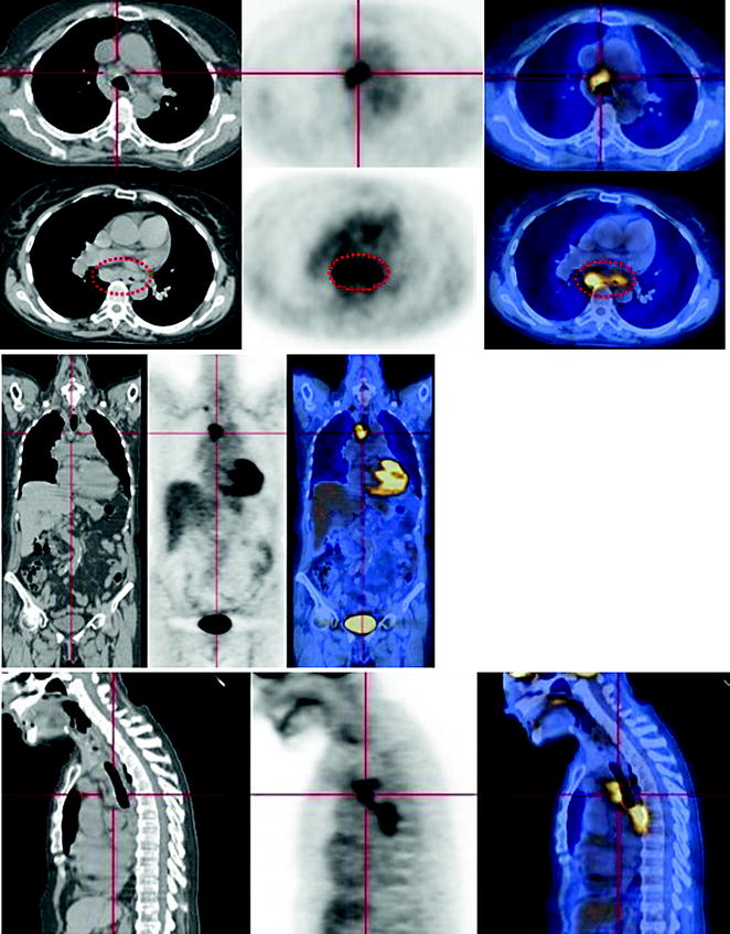
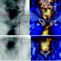
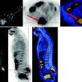
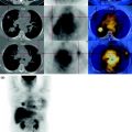
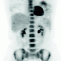
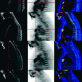
Fig. 22.1
The MIP image shows intense FDG deposition in an esophageal coarse lesion with mediastinal involvement. The use of PET-CT coronal reconstruction allows the definition of the precise location of lymph node metastases in the subcarinal area (arrows)
