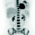
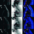
Fig. 21.1
MIP images in anterior and oblique scans show the diffuse high metabolism of the rectum and transverse, descending and sigmoid colon. Caecum and ascending colon show lesser metabolic activity
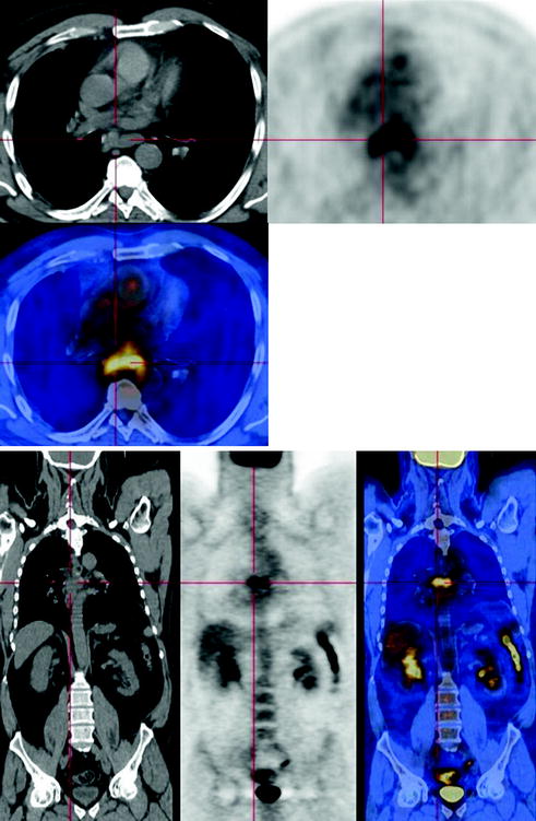
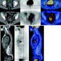
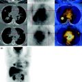
Fig. 21.2


CT-PET shows the presence of solid, inhomogeneous tissue in the posterior mediastinum, inseparable from the wall of the esophagus and showing pathological metabolism
Stay updated, free articles. Join our Telegram channel

Full access? Get Clinical Tree


