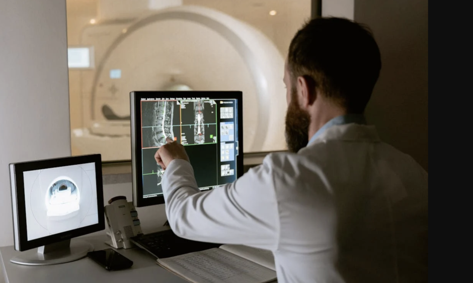
Advanced imaging techniques expose subtle, long-term effects that medications can have on the body. Radiologists are detecting subtle changes linked to long-term drug exposure using advanced imaging techniques.
MRI and CT scans provide detailed views that reveal early signs of damage or abnormal growth. Early identification of medication-related complications remains a challenge in healthcare today.
As chronic medication use rises in people of all ages, what risks might develop silently? How can these imaging tools help doctors balance treatment benefits against potential side effects? What role does imaging play in managing these risks more effectively?
In this article, we will explore how modern imaging reveals the hidden impacts of long-term medication use.
Detecting Skeletal Changes in Patients on Long-Term Therapies
Certain medications reduce bone mineral density and increase fracture risk. Corticosteroids commonly cause weakening of bones with prolonged use. DEXA scans measure bone density and track changes over time.
NIH notes that bisphosphonates can prevent fractures but sometimes cause unusual breaks. Imaging detects these fractures early to guide treatment adjustments. Regular monitoring helps balance medication benefits against bone health risks.
Radiologists interpret scans to advise clinicians on patient management. Bone changes often go unnoticed without imaging surveillance during therapy. Personalized care requires combining imaging results with clinical information.
How does age affect skeletal drug response?
Older patients tend to exhibit faster and more severe bone density decline from chronic treatments. Hormonal changes and decreased physical activity contribute to poorer bone recovery following damage. Regular imaging and proactive bone health strategies become increasingly important as patients age during therapy.
Imaging Methods for Monitoring Liver and Kidney Health
WebMD states that long-term use of some drugs can injure the liver and cause hepatotoxicity. Ultrasound and CT scans identify fatty changes or fibrosis in these organs. Early detection is vital for preventing irreversible damage to vital tissues.
Radiologists look for organ size, texture, and abnormal blood flow patterns. Lab tests sometimes miss early tissue changes that imaging can reveal. Imaging helps doctors adjust medication before severe liver or kidney failure.
Identifying toxicity signs early can improve patients’ quality of life. Cross-sectional imaging is a safe and non-invasive monitoring tool. It supports clinicians in managing complex medication regimens effectively.
Can medications affect one kidney more severely?
Yes, unilateral kidney damage can occur depending on drug distribution, pre-existing asymmetries, or localized conditions. Imaging often reveals differences in size, density, or function between the two kidneys. Recognizing asymmetrical damage helps guide interventions such as targeted therapy cessation or nephrology referral.
Understanding Brain Lesions Linked to Hormonal Agents
Hormonal medications can sometimes affect brain tissue and its functions in unexpected ways. Radiologists have noticed certain tumor patterns that may be linked to hormone exposure. One common type of benign tumor, meningiomas, can grow when stimulated by hormones. These tumors may lead to symptoms like headaches, vision problems, and other neurological issues.
Early detection using contrast-enhanced MRI is crucial for identifying these brain lesions. Often, such tumors are discovered incidentally during scans done for other reasons. Some lesions display characteristics that suggest hormonal influences on their growth.
For example, TorHoerman Law emphasizes that Depo-Provera, an injectable contraceptive containing medroxyprogesterone acetate, has been studied for its potential connection to meningioma development. Prolonged use of this medication may increase the risk of these tumors in certain individuals. Lawsuits have been filed claiming that manufacturers failed to adequately warn users about these risks.
The Depo Provera lawsuit highlights the importance of thorough patient education regarding hormonal therapies. Radiology plays a vital role in detecting these lesions early, which can improve treatment success. Recognizing the imaging features associated with hormone-related tumors helps ensure timely referrals and interventions.
Do genetics influence brain lesion susceptibility?
Yes, genetic predisposition may enhance tissue sensitivity to hormonal changes or medications. Certain polymorphisms affect receptor density or metabolic pathways linked to lesion development. Family history and genetic screening may offer additional insights into individual risk during therapy planning.
Lung Imaging in Patients on Pulmonary Risk Medications
Some medications can cause serious lung damage with long-term use. Radiopedia mentions that amiodarone is a common drug with pulmonary toxicity risks. High-resolution CT scans reveal early lung tissue changes. Radiologists look for ground-glass opacities and fibrotic patterns in the lungs.
Distinguishing drug effects from infections or disease progression is challenging. Regular imaging helps catch damage before severe lung impairment occurs.
Detecting issues early helps adjust or discontinue harmful medications promptly. Radiologic findings guide pulmonologists in managing these patients better. Imaging ensures respiratory health monitoring during risky drug therapies.
Can lung imaging predict future breathing problems?
Yes, patterns like interstitial thickening and ground-glass opacity suggest a high risk for decline. These findings help forecast reduced pulmonary function and future respiratory complications before symptoms arise. Early detection prompts preventive steps like medication adjustment or pulmonary therapy.
Using Imaging to Spot Drug-Related Gastrointestinal Issues
Oxford Academic highlights that long-term use of NSAIDs often causes damage to the gastrointestinal tract. Capsule endoscopy and CT enterography allow detailed visualization of the intestines. Imaging detects ulcers, inflammation, and strictures caused by medications.
Some drugs may induce rare but severe intestinal conditions like ischemia. Radiologists play a key role in linking symptoms to medication side effects. Early identification allows doctors to modify treatment plans accordingly.
Imaging complements clinical exams and laboratory tests for diagnosis. Regular imaging surveillance improves outcomes in patients with GI complaints. Understanding medication effects through imaging enhances patient safety overall.
Can gut changes affect mental health symptoms?
Yes, chronic gastrointestinal inflammation may influence the gut-brain axis, contributing to anxiety or mood changes. Imaging-confirmed damage often correlates with systemic effects beyond digestion, including cognitive and emotional health. Treating gut inflammation may lead to improved psychological well-being over time.
Advanced imaging is changing how we understand the long-term effects of daily medications. It helps doctors look beneath the surface and catch issues before they turn serious. This means care can shift from reacting to problems to preventing them altogether.
Imaging reveals early signs of damage in the brain, bones, lungs, gut, and other organs. That kind of detail lets doctors fine-tune treatments to suit individual needs. When medications are used for years, small changes can matter. These tools give patients and doctors a smarter way to manage risks.
Related posts:
 Coronary Artery Calcium
 Coronary Artery Stents
 General Electric LightSpeed VCT, Optima CT660 and Discovery CT750 HD
 Cardiac MR in Patients with Implantable Arrhythmia Devices
 Top 5 Must-Have Apps for Radiologists in the United States in 2025
How to Improve Radiology Access in Underserved Communities
Top 5 Must-Have Apps for Radiologists in the United States in 2025
How to Improve Radiology Access in Underserved Communities
Stay updated, free articles. Join our Telegram channel

Full access? Get Clinical Tree


