CHAPTER 10 External Auditory Canal Osteoma
Epidemiology
External auditory canal (EAC) osteomas are rare bony outgrowths found in the EAC and are much less common than EAC exostoses. There is no gender predilection and they may be seen in any age group.
Clinical Features
External auditory canal osteomas are most often asymptomatic and detected incidentally. When they are large, they may cause conductive hearing loss. They can also be associated with serous otitis media and cholesteatoma.
Pathology
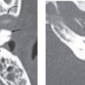
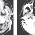
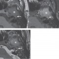
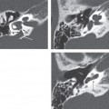
Although EAC osteomas commonly reveal a connection to the underlying EAC bone on gross pathologic examination, in some cases there may be no connection demonstrated. They are most commonly seen at the bony-cartilaginous junction of the EAC. On microscopic examination, they are similar to osteomas seen elsewhere and show irregularly oriented lamellated bone surrounding abundant, discrete fibrovascular channels.
Treatment
Related posts:

Stay updated, free articles. Join our Telegram channel

Full access? Get Clinical Tree


