CHAPTER 4 Facial Nerve
Embryology
The facial nerve is the nerve of the second branchial arch. It serves motor as well as sensory functions. It supplies the striated musculature of the face, neck, and stapedius muscle of the middle ear; parasympathetic fibers to the lacrimal, submandibular, and sublingual glands; the seromucinous glands of the nasal cavity; and it conveys taste sensations from the anterior two thirds of the tongue, the palate, and the tonsillar fossae. It also has a small cutaneous sensory component.
After its origin in the medulla, the facial nerve proceeds forward and laterally in the posterior fossa into the internal auditory meatus, in conjunction with the nervus intermedius (sensory component of the facial nerve) and the acoustic nerve. The intracranial segment of the facial nerve is 23 to 24 mm in length. On entering the internal auditory canal (IAC), the facial nerve occupies the anterosuperior quadrant of this canal for 8 to 10 mm. The internal auditory segment is 7 to 8 mm in length and lies superior to the cochlear nerve, passing above the crista falciformis. While within the canal, the motor root is initially separated from the acoustic bundle by the nervus intermedius, and the two roots unite together in the canal to form the combined trunk.
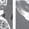
The labyrinthine segment (Fig. 4-1) of the facial nerve measures 3 to 4 mm and represents the narrowest portion of the facial nerve. The facial nerve enters the fallopian canal at the fundus of the IAC and extends from the IAC to the geniculate ganglion. When the nerve reaches a point just lateral and superior to the cochlea, it angles sharply forward, nearly at right angles to the long axis of the petrous temporal, to reach the geniculate ganglion. At the ganglion, the direction of the nerve reverses itself, executing a hairpin bend so that it runs posteriorly. This is the first genu of the facial nerve (Fig. 4-2). The greater superficial petrosal nerve arises from the geniculate ganglion and supplies parasympathetic fibers to the lacrimal gland and seromucinous glands of the nasal cavity (Fig. 4-3).
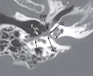
Figure 4-1 The curved reformation of the course of facial nerve nicely demonstrates the various segments of the facial nerve in a single image. The correlative magnetic resonance (MR) scans in the following figures were performed in a patient with Bell palsy that is causing greater than expected enhancement of the facial nerve. However, the enhancement nicely demonstrates the expected location of the nerve in regions of the temporal bone that are often difficult to visualize due to the susceptibility artifact of the surrounding air and adjacent lateral semicircular canal. L, labyrinthine segment; AG, anterior genu; T, tympanic segment; PG, posterior genu; GG, geniculate ganglion; GSP, greater superficial petrosal nerve.
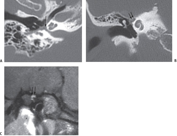
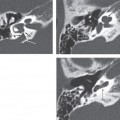
Figure 4-2 (A) Curved reformation of the facial nerve will be used as a localizer for the coronal images through the anterior genu and geniculate ganglion (arrows). (B) Coronal computed tomography (CT) and (C) contrast-enhanced fat-suppressed MR obtained at similar levels through the area of the anterior genu and geniculate ganglion shows the normal location of the nerve at this level (arrows).
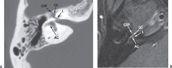
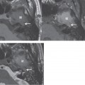
Figure 4-3 (A) Axial CT and (B) contrast-enhanced fat-suppressed MR obtained at similar levels through the epitympanum shows the segmental anatomy of the anterior aspect of the facial nerve. L, labyrinthine segment; AG, anterior genu; GG, geniculate ganglion; GSP, greater superficial petrosal nerve.
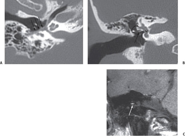
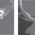
Figure 4-4 Curved reformation of the facial nerve (A) will be used as a localizer for the coronal images through the tympanic segment (arrows). Coronal CT (B) and contrast-enhanced fat-suppressed MR (C) obtained at similar levels shows the location of the facial nerve (arrows
Stay updated, free articles. Join our Telegram channel

Full access? Get Clinical Tree


