Fair Game

Chapter 2 Fair Game


Comment


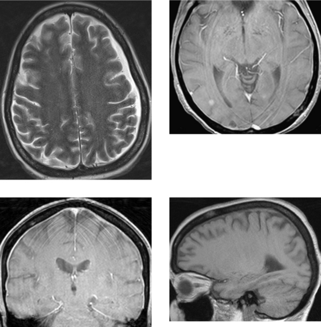
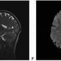
This case of metastatic breast carcinoma illustrates several findings and imaging patterns of metastatic disease. There is a small left subdural effusion that follows the signal characteristics of CSF on all pulse sequences (including FLAIR and diffusion-weighted images, not shown). Note on the axial T2W image that the cortical veins seen as small black flow voids are closely up against the surface of the left cerebrum, consistent with a subdural effusion (in contrast to dilated subarachnoid spaces, as are seen in cerebral atrophy, in which the bridging cortical veins are noted to course through the CSF spaces). There is diffuse thin enhancement of the dura over the cerebral convexities. Axial images also demonstrate two cortical enhancing metastases. Lastly, best appreciated on the sagittal unenhanced T1W image is diffuse decreased signal intensity of the calvarial marrow, with superimposed areas of signal abnormality, consistent with diffuse marrow infiltration or metastases.


Dural metastases usually are secondary to hematogenous dissemination of tumor from extracranial systemic tumors. Dural metastases may also result from direct extension of adjacent calvarial or skull base bone metastases. Breast, lung, and prostate carcinoma as well as melanoma and lymphoma not uncommonly are associated with dural metastases in adults. In children, adrenal neuroblastoma and leukemia are most commonly associated with dural metastases. These malignant neoplasms may also metastasize to the cranial sutures. In infancy, this may present as widened sutures. Inflammatory processes that may mimic dural metastases include granulomatous infections (tuberculosis, syphilis, and fungal), sarcoid, Langerhans’ cell histiocytosis, and Erdheim-Chester disease.


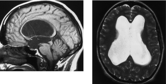
CASE 52 Aqueductal Stenosis


CASE 53 Rathke’s Cleft Cyst


Comment


Rathke’s cleft cysts are embryologic remnants of Rathke’s pouch, the neuroectoderm that ascends from the oral cavity to the sella to form the anterior pituitary lobe and pars intermedia. In the majority of cases, Rathke’s cleft cysts are incidental findings noted at autopsy or identified on imaging studies performed for other reasons. In a recent series of 1000 autopsy specimens, 11% of the pituitary glands had an incidental Rathke’s cleft cyst. These lesions are typically localized within the pituitary sella, although they may also extend into the suprasellar region. In addition, Rathke’s cleft cysts may be centered in the suprasellar cistern anterior to the hypothalamic stalk. A single layer of cuboidal or columnar epithelium, which may contain goblet cells, typically lines these cysts.


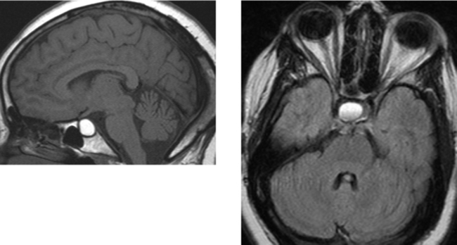
The contents within the cysts are mucoid, resulting in their typical MR imaging appearance. Most Rathke’s cleft cysts are circumscribed and hyperintense on both unenhanced T1W and T2W images. Although this is the most common pattern, cysts may also be hypointense or isointense on either T1W or T2W images, depending on their protein concentration and viscosity. In this case, the cyst is hyperintense on unenhanced T1W imaging and hyperintense on T2W imaging, with a fluid–debris level (did you catch that?). These cysts do not enhance (although there may be mild peripheral enhancement). More recently, intracystic nodules have been described that are hyperintense on unenhanced T1W images and hypointense on T2W images. At surgery, these appear as yellow, waxy solid masses, and the pathologic correlate shows a mucin clump.


Although most Rathke’s cleft cysts are incidental findings, symptoms may occur with larger lesions that compress the optic chiasm (resulting in headache and visual disturbances) or pituitary gland (patients may present with diabetes insipidus or hypopituitarism).


CASE 54 Optic Neuritis (Demyelinating Disease)


Comment


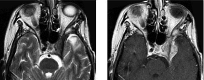
A wide spectrum of disease processes may be associated with optic neuritis, most commonly, demyelinating disease followed by idiopathic disease. Approximately 50% of patients with optic neuritis have multiple sclerosis, and approximately 15% of patients with multiple sclerosis have optic neuritis as their initial clinical presentation. Devic’s disease (neuromyelitis optica) represents a specific syndrome of multiple sclerosis in which patients have isolated transverse myelitis and optic neuritis. These two may present simultaneously or on separate occasions. In older patients, vasculopathies and ischemia are more common causes of optic neuritis.


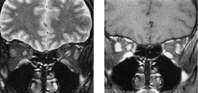
Patients presenting with optic neuritis can be assessed using high-resolution, fat-suppressed, fast spin-echo T2W and enhanced T1W images, as in this case. Imaging in the coronal plane is particularly useful. Imaging may demonstrate increased T2W signal intensity within the nerve itself, and postcontrast images may demonstrate enhancement of the nerve as well as the nerve sheath due to perivenous inflammation (as in this case). In cases of clinically diagnosed optic neuritis, MR imaging of the brain is especially useful and is recommended because it may help to establish the diagnosis of demyelinating disease. In patients with their first episode of optic neuritis, up to 65% have asymptomatic cerebral white matter lesions on brain MR study. In addition, studies have shown that the presence of brain lesions on MR imaging in patients with optic neuritis may be important in determining prognosis and outcome.


CASE 55 Ocular Trauma—Lens Dislocation and Globe Rupture


Comment


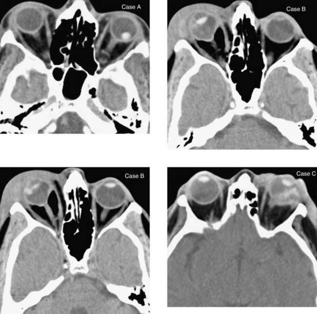
On CT imaging, lens dislocation (Case A) may be differentiated from other intraocular foreign bodies by identifying that the intraocular density has the configuration of a lens and, importantly, observing that the lens in that eye is not located in its normal position. Lens dislocation may be confirmed on physical examination. CT is the imaging modality of choice in the initial assessment of patients with orbital trauma due to its availability as well as the ease and rapidity with which the examination can be performed. In addition, CT readily establishes the presence of orbital foreign bodies, delineates orbital fractures, and assesses for retrobulbar complications of trauma.


Subluxation or dislocation of the lens may be unrelated to trauma. It may be spontaneous or related to infection. There are also hereditary disorders that affect the connective (mesodermal) tissues that may be associated with lens dislocation, such as Marfan syndrome, Ehlers-Danlos syndrome, and homocystinuria. In Marfan syndrome, lens subluxation or dislocation is superior and at the periphery of the globe (and usually bilateral), whereas in homocystinuria, subluxations are inferior (“down and out”).


Globe rupture indicates that the integrity of the outer membranes of the eye is disrupted. Globe rupture may occur when a blunt object impacts the orbit, causing anterior–posterior compression of the globe and raising intraocular pressure such that the sclera tears. Ruptures from blunt trauma usually occur at the sites where the sclera is thinnest, at the insertions of the extraocular muscles, at the limbus, and around the optic nerve. Sharp objects or those traveling at high velocity may perforate the globe directly. Globe rupture represents an ophthalmologic emergency and requires surgical intervention. Damage to the posterior segment of the eye is associated with a very high frequency of permanent visual loss. Early recognition and ophthalmologic intervention are critical to maximizing functional outcome. On CT imaging, the presence of vitreous hemorrhage suggests an associated globe rupture. An enlarged anterior chamber and retraction of the lens are indicative of rupture of the posterior sclera. Other findings of globe rupture are a small, misshapen globe that contains blood or air (as in Cases B and C).


CASE 56 Developmental Venous Anomaly


Comment


Developmental venous anomalies, also referred to as venous malformations or venous angiomas, likely occur late in the first trimester and early in the second trimester of gestation, when the medullary veins are developing. Insults leading to failure in the development of the normal draining venous structures are believed to be the cause of these anatomic variants. Instead of the normal parallel appearance of the medullary veins as they drain into subependymal or superficial cortical veins, a disorganized network of dilated medullary veins converges in a “caput medusa” appearance and drains into an enlarged venous channel that subsequently drains superficially to cortical veins or sinuses, or deeply to subependymal veins of the lateral ventricle and then into the galenic system. Normal cerebral tissue intervenes between the dilated veins. The brain parenchyma surrounding a venous angioma is typically normal, although occasional gliosis, seen as mild T2W hyperintensity, may be present. Developmental venous anomalies are found most commonly in the frontal lobes, followed by the parietal and temporal lobes. Infratentorial developmental venous anomalies occur most commonly in the cerebellum. These can drain to the fourth ventricle and then to the pontomesencephalic vein, or to the precentral cerebellar vein and into the galenic system.


More than 99% of developmental venous malformations are asymptomatic; however, rarely, these lesions may hemorrhage. Hemorrhagic complications are usually secondary to coexisting cavernous malformations. Rare hemorrhage related specifically to the venous angioma is most common with cerebellar lesions. Patients may present with headache, seizure, or focal neurologic symptoms. Asymptomatic (essentially all) developmental venous anomalies are not treated because they represent a compensatory venous drainage route for normal brain. Sacrifice of these lesions could result in venous infarction of the normal brain that they drain.


CASE 57 Fibrous Dysplasia of the Calvaria


Comment


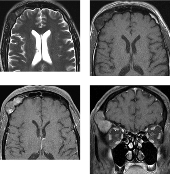
Fibrous dysplasia is a developmental bone disorder in which osteoblasts do not undergo normal differentiation and maturation. The cause is unknown. Fibrous dysplasia may be monostotic (a solitary lesion) or polyostotic (multiple bones involved or multiple lesions in one bone). Approximately 75% of cases of fibrous dysplasia are monostotic (25% polyostotic). Polyostotic fibrous dysplasia involves the skull and facial bones more frequently than does monostotic disease. Common areas of calvarial involvement include the ethmoid, maxillary, frontal, and sphenoid bones. Involvement of these bones may result in orbital abnormalities such as exophthalmos, visual disturbances, and displacement of the globe. Involvement of the temporal bone may result in hearing loss or vestibular dysfunction.


Fibrous dysplasia of the skull or facial bones on plain radiography may present as radiolucent or sclerotic lesions. Sclerotic lesions are more common in the calvaria, skull base, and sphenoid bones. Although Paget’s disease may occur in these same locations of the calvaria, unlike fibrous dysplasia, concomitant involvement of the facial bones is less common. In this case, there is a mass in the right frontal bone, with associated focal expansion of the diploic space. There is preservation of the inner and outer cortical tables without destruction or significant thickening of the cortex. These features are typical of fibrous dysplasia. In contrast, although there may be expansion of the diploic space in patients with Paget’s disease, there is normally thickening of the cortex (which may be extensively involved).


CASE 58 Cavernous Sinus Mass—Meningioma


Comment


A variety of lesions can affect the cavernous sinus, including neoplasms primarily arising from bone (chondrosarcoma, chondroma, and chordoma) and bone metastases. Metastases and lymphoma primarily affecting the cavernous sinus and perineural spread of tumor are also common. Benign neoplasms include meningioma, schwannoma, and extension of a pituitary adenoma. A variety of vascular and inflammatory lesions may also involve the cavernous sinus.


Infectious processes include bacterial and fungal (actinomycosis, aspergillus, and mucormycosis) agents. In addition, the cavernous sinus may be affected indirectly by complications of infectious processes (eg, cavernous sinus thrombosis). Tolosa-Hunt syndrome represents an idiopathic granulomatous inflammatory disorder that may affect the orbital apex and cavernous sinus. Histologically, Tolosa-Hunt syndrome is identical to orbital pseudotumor (differing only in location). It may present with painful ophthalmoplegia, deficits of the cranial nerves coursing through the cavernous sinus, and retro-orbital pain. Symptoms may last for days to weeks, and may be recurrent. Like orbital pseudotumor, it responds rapidly to steroid treatment. MR imaging may show abnormal signal intensity or an enhancing mass within the cavernous sinus. There may be extension into the orbital apex. Thrombosis of the cavernous sinus or superior ophthalmic vein may occur.


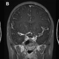
These images demonstrate a mass within the left cavernous sinus that is hypointense to CSF (isointense to brain parenchyma) on T2W imaging. Neoplastic processes that may be hypointense on T2W imaging include lymphoma, meningioma, and less commonly, plasmacytoma and schwannoma. Sarcoid may also be hypointense on T2W imaging. Enhancement extends along the lateral dural margin, as well as posteriorly along the tentorial margin (an appearance that may be seen with meningiomas, lymphoma, and sarcoid). The marked narrowing of the left cavernous internal carotid artery with preserved antegrade flow makes meningioma the best diagnosis. Biopsy through a left temporal craniotomy revealed a meningioma.


CASE 59 Pyogenic Ventriculitis with Acute Hydrocephalus


Comment


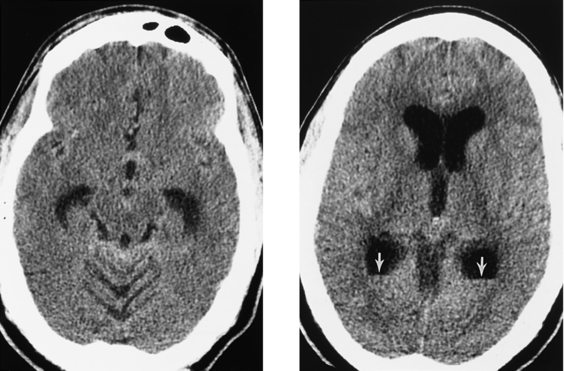
This case demonstrates acute hydrocephalus and fluid–fluid levels (arrows) within the occipital horns of the lateral ventricles, consistent with ventriculitis complicating pyogenic meningitis. Lumbar puncture yielded pus. Ventriculitis is due to the introduction of infectious organisms into the ependyma or ventricles, and may be secondary to bacteremia, extension of an intraparenchymal abscess, trauma, or surgical instrumentation (especially placement of ventricular shunts). In addition to ventriculitis, other complications of ventriculoperitoneal shunts include overdrainage resulting in slit ventricle syndrome, subdural hematomas (which are frequently bilateral), shunt malfunction, surgery-related complications, and metastases in the setting of neoplasm (such as primitive neuroectodermal tumor).


Approximately 20% of patients with pyogenic bacterial meningitis will have complications necessitating neurosurgical intervention (surgery or a ventriculostomy), even after antibiotic therapy. Such complications include subdural empyema; parenchymal brain abscess; ventriculitis with hydrocephalus, as in this case; and encephalitis. The development of such complications may correlate with inadequate treatment or with the duration of meningitis before the initiation of therapy.


Cytomegalovirus (CMV) ventriculitis is unusual, but may occur in patients with HIV infection. Most HIV-infected patients with CMV ventriculitis have already been diagnosed with an AIDS-defining condition. Pathologic findings include inflammation of the ependyma and periventricular structures, as well as ependymal necrosis with CMV intranuclear inclusion bodies. The differential diagnosis in this patient population includes non-Hodgkin’s lymphoma.


CASE 60 Lesions of the Pituitary Stalk and Hypothalamus: Case A—Sarcoidosis; Case B—Langerhans’ Cell Histiocytosis


Comment


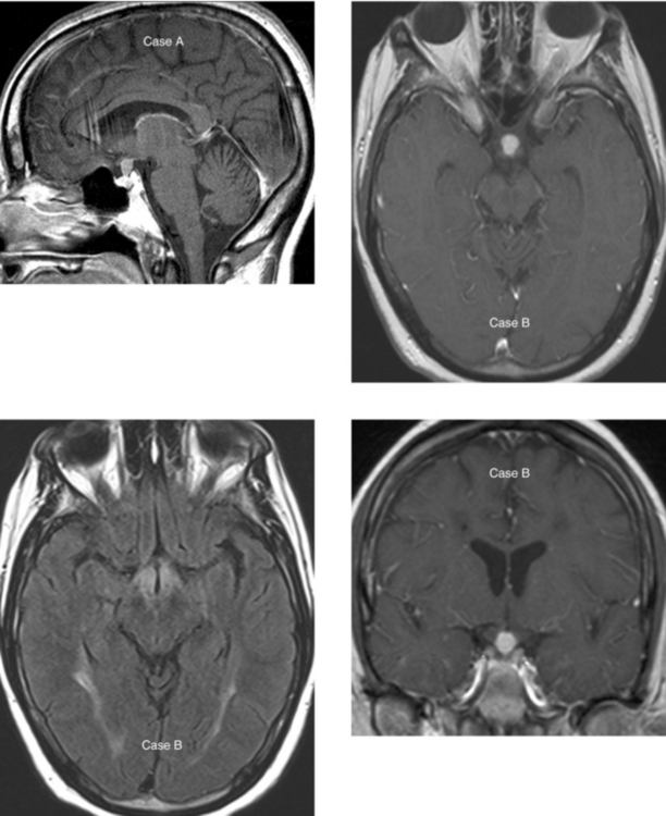
In Case A, the midline sagittal enhanced T1W MR image shows abnormal thickening and enhancement along the pituitary stalk that lacks normal tapering as it extends inferiorly toward the pituitary gland. This represented sarcoid, and responded well to steroid treatment. This patient had a history of sarcoid with pulmonary involvement. Involvement of the CNS by sarcoid occurs in approximately 5% to 15% of patients, with isolated involvement of the pituitary gland or stalk in fewer than 1% to 2% of all patients with sarcoid. When CNS sarcoid presents with leptomeningeal involvement, the pituitary stalk in these cases is commonly involved.


Case B shows an enhancing mass in the hypothalamic region at the level of the upper pituitary stalk, and this represented Langerhans’ cell histiocytosis. The differential diagnosis of an infundibular mass in children includes germinoma, infection (meningitis or tuberculosis) and, less commonly, lymphoma or leukemia and glioma involving the hypothalamic region. In patients with Langerhans’ cell histiocytosis, there may be absence of the normal “bright spot” of the posterior pituitary gland in the sella on unenhanced T1W images. Langerhans’ cell histiocytosis uncommonly affects the CNS. It is a disorder of the reticuloendothelial system. The most common cranial bone abnormality is a lytic, circumscribed, enhancing mass of the calvaria or skull base. In the cranium, it most commonly involves the temporal bone along the mastoid segment. It also may involve the frontal bone along the orbit.


The differential diagnosis of an infundibular lesion in adults includes granulomatous disease (sarcoid or tuberculosis) and metastasis (especially lung and breast). Less commonly, lymphoma or a hypothalamic glioma may present in this way. Lymphocytic adenohypophysitis is a unique condition that almost always affects women. It is most common in the postpartum period or in the late stages (third trimester) of pregnancy. It is characterized by lymphocytic infiltration of the adenohypophysis and is usually self-limited.


CASE 61 Lhermitte-Duclos Disease (Dysplastic Gangliocytoma of the Cerebellum)


Comment


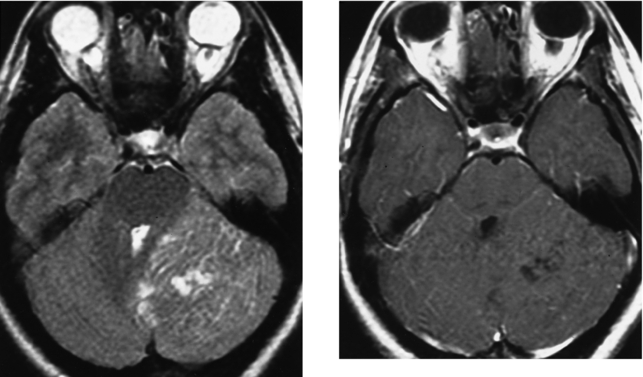
Lhermitte-Duclos disease is also known as dysplastic gangliocytoma of the cerebellum. When symptomatic, patient typically presents with complaints related to mass effect in the second and third decades of life. Lhermitte-Duclos disease has been associated with Cowden disease (multiple hamartoma syndrome), an autosomal dominant disorder associated with an increased incidence of neoplasms in the pelvis, breast, colon, and thyroid. Intracranial meningiomas have also been noted with this syndrome.


Although controversial, Lhermitte-Duclos disease is considered to represent a complex hamartomatous malformation, not a true neoplasm. Dysplastic gangliocytomas often occur in the cerebellar hemispheres. These lesions tend to be poorly demarcated, presenting on CT as a mildly hypodense mass. Calcification has been reported. On MR imaging, these lesions have a more characteristic appearance in which the gray and white matter of the cerebellar hemisphere are both involved and are thickened and hyperintense on T2W imaging, showing a somewhat characteristic laminated or “corduroy” appearance. They may exert mass effect. Hydrocephalus may be present. Dysplastic gangliocytomas do not demonstrate significant enhancement. On pathologic evaluation they usually appear as dysplasia with cellular disorganization of the normal laminar structure of the cerebellum, and hypertrophied granular cell neurons. Histologically there is hypermyelination of axons, and pleomorphic ganglion cells replace the granular and Purkinje cell layers.


CASE 62 Basilar Meningitis—Sarcoidosis


Comment


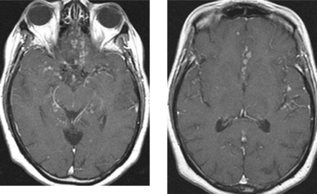
Sarcoidosis is a systemic disorder characterized pathologically by noncaseating granulomas. Typical presentation is in the third and fourth decades of life, and it is slightly more common in women than in men. CNS involvement has been reported in 5% to 15% of cases, and isolated CNS involvement occurs in fewer than 2% to 4% of cases. Multiple patterns of CNS involvement may occur. The most common is chronic meningitis with a predilection for the leptomeninges of the basal cisterns, as in this case. These patients may present with chronic meningeal symptoms, cranial neuropathies (especially involving the facial and optic nerves), or symptoms related to involvement of the hypothalamus and pituitary stalk. Imaging findings are best demonstrated with MR imaging and include nodular enhancement of the leptomeninges that tends to be most pronounced in the basal cisterns (but may be seen in any of the subarachnoid spaces, as in this case). Another common pattern of CNS sarcoid is brain involvement, with multiple regions of abnormality that may occur as a result of direct extension from leptomeningeal disease; white matter lesions mimicking multiple sclerosis may be present because of disease extension along the perivascular spaces or related to sarcoid-induced small vessel vasculitis. There may also be granulomas in the brain. Less commonly, dural-based disease may be the predominant imaging finding and may be mistaken for a meningioma.


Central nervous system tuberculosis may have a variety of clinical and radiologic presentations, including nodular tuberculous meningitis similar to sarcoid, cerebritis, abscess formation, tuberculoma, or a combination of these. CNS tuberculosis is normally related to hematogenous dissemination from a systemic source, such as the lung, genitourinary system, or gastrointestinal tract. Arteritis may be seen in up to one third of patients with basilar meningitis. This is because the vessels coursing through this inflammatory exudate may become directly involved. Consequences of arteritis include vasospasm and infarction.


CASE 63 Pituitary Microadenoma


Comment


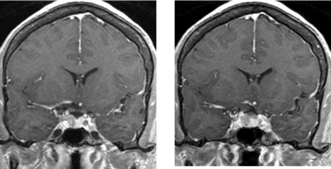
Pituitary adenomas are slow-growing, benign epithelial neoplasms arising from the anterior lobe of the gland. They are typically demarcated with a “pseudocapsule” that separates them from the normal gland. Pituitary adenomas larger than 10 mm are referred to as macroadenomas, and those less than 10 mm in diameter are referred to as microadenomas. Many adenomas are incidental findings (asymptomatic).


The clinical presentation of pituitary adenomas depends on their size, the presence of hormone secretion resulting in endocrine hyperfunction, and the presence of extension beyond the sella (leading to visual symptoms or cranial nerve palsies related to compression). In vivo, approximately 75% of pituitary adenomas are hormonally active (however, in autopsy series, nonsecreting adenomas are much more common).


The most common clinically significant secreting adenoma is the prolactinoma (which arises from the prolactin-secreting cells [lactotrophs]). In women, the most common clinical presentation is irregular menses, galactorrhea, and infertility. In men, impotence may be present. Hormonally active pituitary adenomas arising from somatotrophs, the growth hormone-secreting cells, cause acromegaly in adults and gigantism in children. In acromegaly, endochondral and periosteal bone formation is stimulated, as is proliferation of connective tissue. These changes result in bone overgrowth and increased soft tissue thickness, especially in the “acral” regions (feet, hands, mandible). There is enlargement of the mandible, thickening of the cranial vault, and frontal bossing (the result of enlargement of the frontal sinuses and prominence of the supraorbital ridges).


CASE 64 Multiple Sclerosis—Marburg Type


Comment


Although the etiology of multiple sclerosis is unknown, several causative factors have been implicated. These include autoimmune disease, infection (viral agent), and genetic factors. The prevalence of multiple sclerosis varies with geographic location.


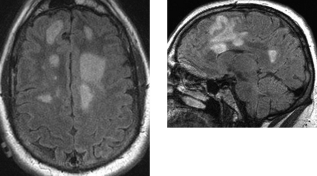
Variants of multiple sclerosis may be present on a clinical or imaging basis. A handful of rare borderline types of multiple sclerosis occur, including Marburg type (also known as acute, fulminant, or malignant multiple sclerosis), a form of acute multiple sclerosis usually seen in younger patients that may be preceded by fevers, is typically rapidly progressive, and can result in death. In such cases, there is extensive demyelination and there may be defined rings within or surrounding plaques of acute demyelination. Enhancement is typically seen in the region of these rings. Concentric sclerosis or Balo-type sclerosis is characterized histologically by alternating rings of demyelination and myelination (normal brain or areas of remyelination) and has a characteristic MR imaging appearance. Schilder’s disease is a rare progressive demyelinating disorder that usually begins in childhood. Symptoms may include dementia, aphasia, seizures, personality changes, tremors, balance instability, incontinence, muscle weakness, headache, and visual impairment.


A type of multiple sclerosis that is usually limited to the optic nerves and spinal cord (either simultaneously or separately) is Devic’s disease or neuromyelitis optica. The main symptoms of Devic’s disease are loss of vision and spinal cord dysfunction. The visual impairment can consist of reduced visual fields, diminished light sensitivity, or loss of color vision. Spinal cord dysfunction includes muscle weakness and lack of coordination, reduced sensation, and incontinence. The brain is usually spared.


“Tumefactive” multiple sclerosis on imaging may be mistaken for a neoplasm or occasionally an abscess, particularly in the absence of a clinical history. The age of the patient may be helpful (multiple sclerosis typically occurs in younger patients). In addition, on close questioning, patients often have neurologic symptoms that are spaced in both time and location. MR imaging may show white matter lesions separate from the mass, suggesting multiple sclerosis. Unlike neoplasms, tumefactive multiple sclerosis often has relatively little mass effect for the amount of signal abnormality present.


CASE 65 Transtentorial Herniation


Comment


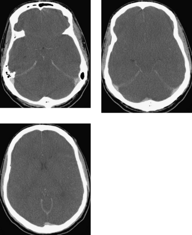
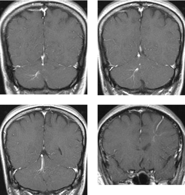
This case illustrates global cerebral swelling after resuscitation from a cardiac arrest resulting in transtentorial herniation. The brain is confined by the cranial vault and an inelastic dura. CSF surrounding the brain serves as a shock absorber. With increased intracranial pressure, herniation of brain from one compartment to another may occur. There are multiple patterns of herniation. In the posterior fossa, there may be inferior tonsillar and cerebellar herniation, with displacement of these structures into the foramen magnum, or there may be upward herniation (superior vermian herniation) in which cerebellar tissue obliterates the quadrigeminal and superior vermian cisterns. Herniation syndromes in the supratentorial compartment include subfalcine, temporal lobe, and central transtentorial herniation.


Transtentorial herniation is typically caused by supratentorial mass lesions or global cerebral swelling, resulting in vector forces that are directed medially and inferiorly. There is resultant effacement of the basilar cisterns and ventricles, as well as caudal displacement of the upper brainstem. The temporal lobe shifts over the tentorium. Caudal displacement of the brainstem and effacement of the ambient cisterns may result in compression of the oculomotor nerve (resulting in ipsilateral pupillary dilation) and compression of the posterior cerebral arteries, resulting in ischemic changes in the occipital lobes and brainstem or diencephalon. There may be secondary small hemorrhages (Duret), which are typically centrally located within the rostral pons and midbrain. Other findings include pseudosubarachnoid hemorrhage and pseudovenous sinus thrombosis, in which the subarachnoid spaces and venous sinuses appear hyerdense on unenhanced imaging relative to the diffuse hypodensity of the edematous cerebrum, as in this case.


Jan 22, 2016 | Posted by in NEUROLOGICAL IMAGING | Comments Off on Fair Game

Full access? Get Clinical Tree

Get Clinical Tree app for offline access