Fair Game

Chapter 2 Fair Game


Aneurysmal Bone Cyst (C2)


Comment


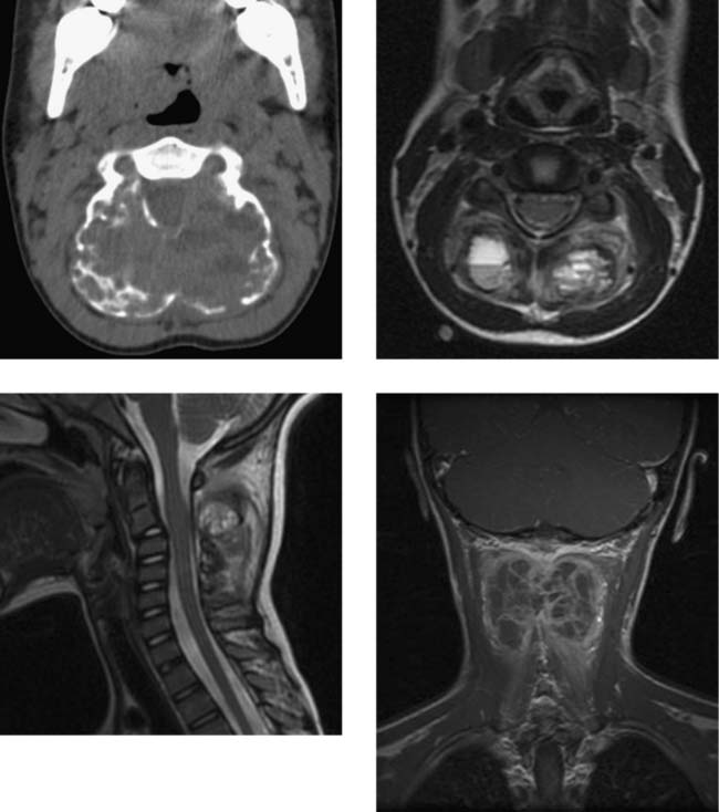
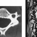
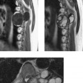
Aneurysmal bone cyst is a benign, highly vascular osseous lesion of unknown origin. It accounts for approximately 1% to 2% of primary bone tumors, and 3% to 20% of ABCs involve the spine. Upper cervical lesions are rare. Patients with ABC are usually in the second decade of life. The pedicles and posterior elements are involved in most cases. In this C2 lesion, the bilateral pars interarticularis, facets, and posterior arch are involved by the expansile lesion, resulting in a kyphosis at C2–C3 (see midsagittal image). Cord compression at C2–C3 (shown on CT and midsagittal MR images) is likely due to vertebral expansion as well as epidural extension of the cystic components of the lesion through minor fractures in the thin cortical bone. Histologically, the interior of the ABC can be solid and vascular and/or cystic and hemorrhagic (surrounded by a thin bony rim). CT demonstrates the osseous expansion and can detect the multiple small fluid levels, although these are more evident on axial and sagittal T2W MR images. Within the cysts, the dependent fluid is hypointense, whereas the nondependent fluid is hyperintense as a result of the different sedimentation properties and composition of blood products in the two components. The internal septations and the peripheral tissue of the ABC are reported to enhance smoothly on postcontrast T1W images (see coronal image), potentially distinguishing ABC from neoplastic lesions, which are more likely to exhibit nodular enhancement.


Notes


Dural Arteriovenous Fistula


Comment


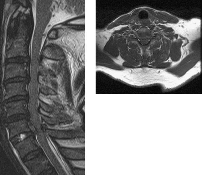
The findings of cord signal abnormality and enhancement are nonspecific and may result from neoplastic, inflammatory, or vascular conditions. Cord enlargement usually favors neoplasm although any intramedullary process that produces edema and breakdown of the blood-cord barrier may mimic neoplasm. The finding of multiple “flow voids” is the key to narrowing the differential diagnosis. Their presence favors a diagnosis of vascular malformation or vascular tumor although craniospinal hypotension or collateral venous drainage secondary to inferior vena cava occlusion can produce enlargement of intradural veins. In this case, no intramedullary tangle of vessels was identified. Thus, an intramedullary arteriovenous (AV) malformation causing the perimedullary flow voids was considered less likely than a dural AV fistula. Vascular tumor, such as hemangioblastoma, was also unlikely, since no focal nodular or mass-like enhancement and no associated cord cyst were detected.


For dural AV fistulas, abnormal MR findings are observed with approximately the following frequency: cord hyperintensity on T2W images, 90% to 100%; cord enlargement, 60% to 70%; cord enhancement (patchy or diffuse) with gadolinium, 60% to 90%; and abnormal intradural (subarachnoid) vessels, 40% to 80%. Cord enhancement results initially from spinal venous hypertension caused by the retrograde flow of blood from the fistula into the veins surrounding and within the cord. Without treatment, venous infarction develops over time.


Notes


Sickle Cell Disease with Osteomyelitis and Epidural Abscess, Lumbar


Comment


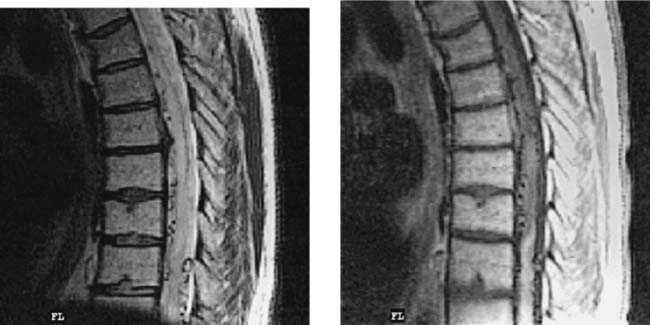
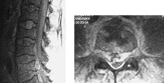
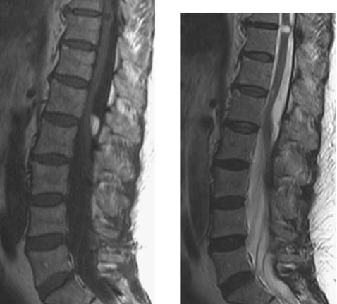
Sickle cell patients are more prone than others to certain infections, including pneumococcal and Salmonella infections, as a result of decreased phagocytic ability of the reticuloendothelial system. This patient had Streptococcus pneumoniae sepsis and meningitis. She had been treated with antibiotics for 12 days when MR images were obtained because of persistent fever and back pain. The sagittal proton density FSE image, covering the T12–L1 to L4–L5 disk spaces, shows diffusely decreased signal intensity within the vertebral bodies relative to the disk spaces, consistent with bone marrow hyperplasia. The body of L4 is slightly hyperintense in comparison with the other vertebral bodies, and its inferior endplate is indistinct—features suggestive of osteomyelitis. The adjacent disk space had a normal appearance on all other sequences. A variably hyperintense anterior epidural collection extends from L3 through L5, with marked compression of the thecal sac and cauda equina at L4. The postcontrast, fat-saturated T1W axial image at L4 demonstrates heterogeneous enhancement within the L4 body and in the paravertebral and epidural spaces. An anterior epidural abscess was confirmed at surgery.


The skeletal imaging findings in sickle cell disease are due to (1) bone marrow hyperplasia secondary to anemia and (2) bone and marrow ischemia and infarction secondary to sickling episodes. In the spine the vertebral bodies have been described as having an H shape and/or a smoothly curved endplate depression. The H vertebral body shape has been attributed to a growth disturbance of the central area secondary to ischemia/infarction. Some authors distinguish this shape from a smoothly curved endplate depression attributed to osteoporosis secondary to bone marrow hyperplasia. Other authors use the general term biconcave to refer to the vertebral endplate contour abnormalities.


Notes


Chordoma, Sacral


Comment


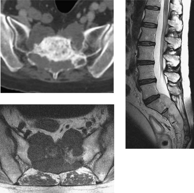
The CT and MR images show a soft tissue mass that appears to originate in the sacrum from S1 to S3, causing bone destruction. The mass is midline and extends into the sacral ala and the presacral region as well as the sacral canal. On the sagittal T2W image, the mass is hyperintense. The findings are typical for a destructive neoplasm but are not specific, and several tumors may be considered in the differential diagnosis. Two findings may help distinguish chordoma from other neoplasms: (1) a midline location (as shown here) and (2) the appearance on T2W images of hypointense septations separating hyperintense lobules that make up the bulk of the mass.


Because they originate from notochordal remnants, chordomas may involve any segment of the craniospinal axis from the sphenoid to the coccyx. The approximate frequency of involvement is as follows: sacrococcygeal, 50%; clival, 35%; remainder of the spine, 15%. Sacral chordomas usually extend anteriorly rather than posteriorly, so that they compress or invade the lower sacral nerves. Typical chordomas consist of mucin-containing physaliphorous cells and/or purely cystic mucinous pools. These account for the hyperintense lobulations of the mass that are seen on T2W images. Hypointense septations represent fibrous strands between the lobular components of the tumor. Sacral chordoma occurs with almost twice the frequency in men as in women. It is uncommon in individuals younger than 40 years of age. Chordomas are typically slow growing but are locally aggressive, and gluteal muscle infiltration due to lateral extension of tumor is common. Most sacral chordomas displace rather than invade the rectum. Metastasis is usually a late event. The presenting symptom in the vast majority of patients is local pain. Approximately one third of patients also have radiculopathy due to irritation of the sciatic nerve or iliolumbar trunk.


Notes


Multiple Sclerosis, Cervical


Comment


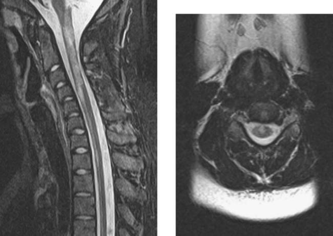
The sagittal image demonstrates diffuse, heterogeneous hyperintensity and mild enlargement of the upper cervical cord, as well as anterior cord involvement at and below C4–C5. On the axial image, the anterior cord involvement appears to encompass the anterior median sulcus and the region of the anterior columns. Although there also appears to be some involvement of the central gray matter, raising the possibility of anterior spinal artery (and/or branches) ischemia/infarction, the characteristic findings of a double-dot or H-shaped hyperintensity in the central gray matter due to infarction are not observed. In a study of cervical and thoracic, predominantly short-segment MS plaques, each occupying less than 50% of the cord cross-sectional area, Tartaglino et al found that the epicenter for the plaques was anterior/anterolateral cord periphery in 20% and posterior/posterolateral cord periphery in 79%. Only 1% of plaques had a central location.


The white matter tracts in the spinal cord are predominantly longitudinally oriented structures that more or less restrict the movement of water molecules depending on the direction of their average displacement. Thus, water molecules that diffuse parallel to the long axis of the tracts (i.e., in approximately the superior-inferior direction) essentially move without restriction, whereas molecules that diffuse perpendicular to the long axis (i.e., in the right-left or anteroposterior direction) have restricted motion. This phenomenon is referred to as diffusion anisotropy. If the fiber structure of the white matter is disrupted, for example, by demyelination and especially axonal loss, diffusion perpendicular to the long axis of the fibers becomes less restricted, and anisotropy is diminished. In addition, less restricted motion in the right-left and/or anteroposterior directions results in an increase in the mean diffusivity. Preliminary reports indicate that such changes are occurring in chronic MS plaques of the spinal cord. For acute inflammatory lesions, edema may contribute to the increased mean diffusivity and the loss of anisotropy. Measures of white matter integrity based on MR diffusion imaging may prove to be more sensitive than standard spin-echo imaging of cord morphology based on T1 and T2 relaxation times and spin density of water protons. Preliminary studies suggest that acute spinal cord infarction, like acute brain infarction, results in decreased mean diffusivity (equivalent to the trace of the apparent diffusion coefficient, or ADC).


Notes


Vertebral Lymphoma with Epidural Venous Enlargement


Comment


The left parasagittal fat-saturated T1W image demonstrates a pathologic fracture of T11 with abnormal enhancement posteriorly in the bodies of T10 and T11. Retropulsion of the T11 body was shown on adjacent images. The confluent epidural enhancement follows the course of the left anterior internal vertebral vein from T11 to the lumbar region. The axial image at the T12 level shows that both anterior internal epidural veins are enlarged. The venous engorgement may be explained as follows: (1) compression of the anterior venous plexus by tumor or displaced bone at T11 and/or (2) intravenous tumor or thrombosis at T11. Biopsy of T11 revealed B-cell lymphoma. The question then arises about whether the posteriorly located lesion in the body at T10 is the result of local spread of lymphoma from T11 via the vertebral venous plexuses.


The vertebral venous system is a valveless anastomotic system in which blood flows either cephalad or caudad, depending on changes in intrathoracic and intra-abdominal pressure and postural factors. The anterior internal vertebral venous plexus has a relatively constant morphologic pattern consisting of a pair of longitudinal trunks each located anterolaterally in the canal. The trunks deviate laterally at the level of the intervertebral disk and converge medially at the midpoint of the vertebral body, where they unite with the basivertebral vein. The trunks are located anterior to the PLL at the level of the vertebral body and then posterior to the PLL where it merges with the intervertebral disk. Compared with the anterior internal plexus, the posterior internal plexus is smaller, less constant, and more difficult to opacify on radiologic studies. Anastomoses exist between the anterior and posterior internal plexuses at each vertebral level and between the internal and external venous plexuses.


Notes


Intradural Lipoma, Lumbar


Traumatic Disk Herniation, Cervical


Metastasis Mimicking Diskitis/Osteomyelitis


CNS Dissemination of Conus Ependymoma


Comment


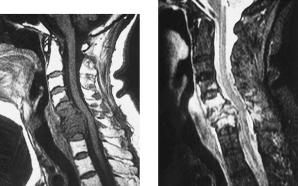
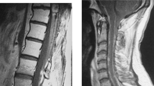
The postcontrast image of the thoracolumbar spine shows evidence of surgery with laminectomies from T10–T11 to L1, inhomogeneous enhancement of the cauda equina and conus, and a residual conus mass (upper edge of the figure). The bodies of T11–L2 demonstrate marked hyperintensity, compatible with postradiation changes. The postcontrast image of the cervical spine, obtained 1 year later, demonstrates leptomeningeal enhancement, with multiple nodular foci extending from the cervical region to the basal cisterns of the posterior fossa and tonsillar ectopia. No irradiation changes are detected in the cervical vertebrae. The combination of findings on the two images suggests that this patient has leptomeningeal metastatic disease secondary to CSF spread of metastases from a conus tumor (most commonly an ependymoma) that was incompletely resected and that this patient received postoperative radiation therapy.


The incidence of dissemination of primary spinal ependymomas is 12.5%, versus 9.6% for primary intracranial ependymomas. Most of the disseminated tumors from primary spinal lesions are of the myxopapillary histologic subtype. This subtype is commonly found in primary ependymomas of the filum terminale, cauda equina, or conus (as in this case). The goal of surgical treatment of both intracranial and spinal ependymomas is gross total resection. The frequency of CNS dissemination following surgery is strongly dependent on the extent (total vs. subtotal) of the initial resection.


Notes

Stay updated, free articles. Join our Telegram channel

Feb 14, 2016 | Posted by in MUSCULOSKELETAL IMAGING | Comments Off on Fair Game

Full access? Get Clinical Tree
Get Clinical Tree app for offline access