For the radiologist and the orthopedic surgeon, the “fall from standing height” is perhaps the most common etiology seen on the daily worklist. While the imaging of a femoral neck fracture is routine, the mechanics of the fall itself (specifically those occurring during bed egress) represent a critical area for preventative intervention. Furthermore, the role of patient positioning extends beyond trauma, serving as the primary defense against the deep tissue pathologies that necessitate complex imaging.
The epidemiology of geriatric trauma is dominated by the fall. According to data from the CDC, falls are the leading cause of injury-related death among adults age 65 and older. For the radiology department, this translates into a constant, relentless stream of pelvic X-rays, hip CTs and MRI scans to detect occult fractures.
However, a significant percentage of these falls do not occur while the patient is ambulating through a community or navigating icy pavement. They occur at the bedside, specifically during the transition from the supine to the standing position. This “egress” phase is biomechanically demanding, requiring a coordination of musculoskeletal strength, proprioception and cardiovascular stability that many geriatric patients simply no longer possess.
Understanding the physics of this transfer (and the equipment that can mitigate its risks) is essential for reducing the radiologic volume of acute trauma cases. Yet, the utility of advanced medical equipment extends beyond fall prevention. It is also the first line of defense against the chronic immobility that leads to osteomyelitis, a pathology that frequently challenges the diagnostic acumen of the radiologist.
The Biomechanics of the Sit-to-Stand Transfer
The “Sit-to-Stand” (STS) cycle is one of the most mechanically complex daily activities. It requires the patient to translate their Center of Mass (COM) forward from a stable seated position to an unstable bipedal stance.
For a healthy adult, this involves generating significant flexion momentum at the hip, followed by rapid extension of the knee and hip joints. It is a precise kinetic symphony. However, in the geriatric population, this kinetic chain is often compromised. Sarcopenia (age-related muscle loss) specifically targets the quadriceps and gluteal muscles required for this lift.
Furthermore, the transition often triggers orthostatic hypotension: a sudden drop in systolic blood pressure upon standing. If a patient attempts to egress unassisted, the combination of muscular weakness and transient cerebral hypoperfusion creates a high-risk window for a collapse. This is the moment where the femoral neck is most vulnerable to the direct impact forces that necessitate urgent radiographic evaluation.
The “pre-egress” phase is equally critical. Before a patient can even attempt to stand, they must rotate their body 90 degrees on the mattress and drop their legs over the side. In a standard static bed, this is a high-friction maneuver. The elderly patient must perform a “scoot and twist,” introducing significant shear force on the skin and rotational torque on the lumbar spine. It is during this unassisted struggle against friction that many patients slip or lose their balance before their feet even touch the floor.
The “Osteomyelitis” Link: Preventing Deep Tissue Pathology
While acute trauma from falls is the most visible consequence of poor bed mobility, there is a slower, more insidious pathology that radiologists frequently encounter: Osteomyelitis secondary to pressure ulcers.
Immobility is the enemy of tissue viability. When an elderly patient cannot easily exit the bed or reposition themselves, they are at high risk for decubitus ulcers. As these ulcers progress from Stage 1 to Stage 4, they penetrate the fascia and expose the underlying osseous structures.
For the radiologist, diagnosing osteomyelitis in this context is complex. Plain films may show cortical erosion or periosteal reaction, but often only in late stages. MRI is the gold standard, often revealing marrow edema and soft tissue abscesses. However, once the pathology has reached the point of requiring an MRI, the patient is often facing surgical debridement or amputation.
The most effective “treatment” for these radiological findings is prevention through mobilization. This is where the mechanics of the bed become a clinical tool for infection control. A bed that assists in rotation allows for more frequent, less painful repositioning. It encourages the patient to get out of bed more often, restoring perfusion to tissues that would otherwise be compressed. By facilitating movement, we prevent the soft tissue breakdown that eventually allows bacteria to invade the bone marrow, effectively reducing the caseload of complex infectious disease imaging.
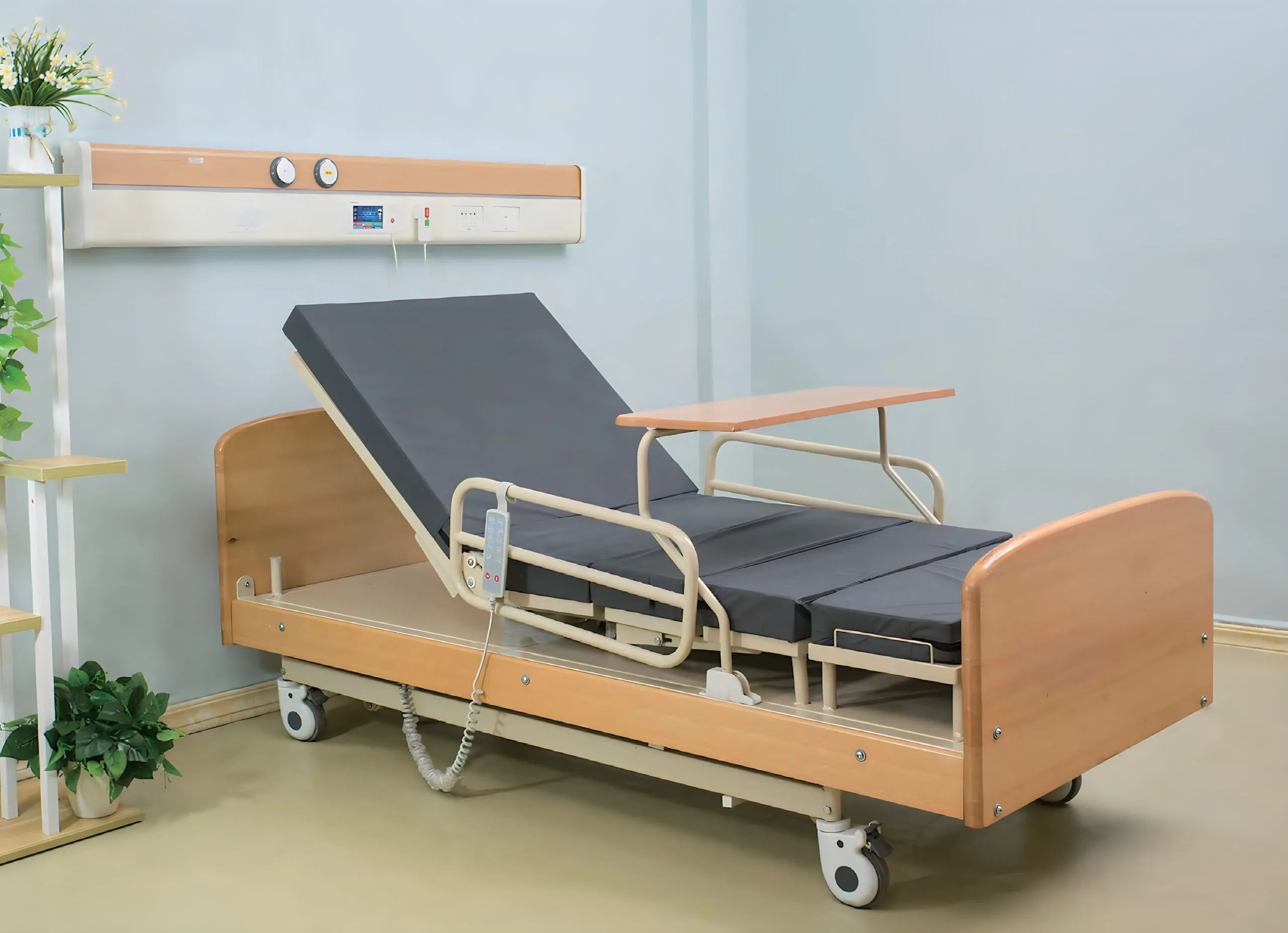
Engineering Safety: The Role of Rotating Hospital Beds
Standard hospital beds are static platforms. They allow for elevation of the head and feet (Trendelenburg positioning), but they do not actively assist in the lateral biomechanical rotation required for egress. This forces the patient to rely entirely on their own compromised core strength.
To mitigate this, clinical protocols are increasingly adopting dynamic support surfaces. Specialized rotating hospital beds for elderly patients are designed to mechanize the most dangerous phase of the transfer. These beds can rotate the mattress 90 degrees and tilt the patient into a seated position with their feet touching the floor, all while the patient remains supported by the bed structure.
By passively orienting the patient toward the egress point, the equipment effectively eliminates the need for the dangerous rotational torque. It transforms the patient from a passive load into an active participant in a controlled “stand assist” maneuver. This drastic reduction in biomechanical demand allows patients with severe sarcopenia or neurological deficits (such as post-stroke hemiparesis) to exit the bed without the “dead weight” struggle that precipitates a fall.
Radiological Implications of Fall Prevention
From an imaging perspective, the goal of deploying such equipment is to reduce the incidence of high-velocity impact injuries.
As detailed in radiological literature regarding femoral neck fractures, the pathology is often determined by the nature of the fall. A lateral fall from a standing height directs energy through the greater trochanter. In osteoporotic bone, this force is catastrophic. It often results in complex intertrochanteric or subcapital fractures that require internal fixation or hemiarthroplasty.
The vector of the fall matters. Falls that occur during the “twisting” motion of getting out of bed often result in spiral fractures of the femur or compression fractures of the spine due to the awkward angle of impact.
By stabilizing the egress process, clinicians can prevent the specific vector of falls that results in these high-acuity fractures. A patient who is supported by a rotating mechanism during the transition to standing is far less likely to suffer the uncontrolled lateral collapse that fills the trauma bays. Thus, the investment in biomechanically appropriate durable medical equipment (DME) has a direct inverse correlation with the utilization of emergency imaging services. The best radiograph is the one that never needs to be ordered.
Discharge Planning and Home Environment
The challenge of safe egress does not end at the hospital door. With the rise of “Hospital-at-Home” models and the push for earlier discharge to reduce nosocomial infection risks, patients are being sent home with higher acuity levels than in previous decades.
For the discharge planner and the family, replicating the safety of the ward environment is the primary hurdle. A patient who requires “Maximum Assist x2” (two people to help) to get out of bed in the hospital cannot be safely discharged to a home where they live alone or with a frail partner.
This is where the prescription of rotating hospital beds becomes a vital component of the discharge plan. It allows for a single caregiver (or even the patient independently) to manage the STS transfer safely. It bridges the gap between the clinical environment and the domestic one.
Furthermore, it addresses the “fear of falling” (post-fall syndrome). Many patients, after suffering a fracture, develop a psychological paralysis. They are afraid to move, which leads to muscle atrophy, which increases the risk of another fall. The security provided by a mechanized chair-bed restores confidence. It allows the patient to mobilize frequently, maintaining bone density and proprioception.
A New Standard for Patient Safety
The intersection of radiology and durable medical equipment is often overlooked. Radiologists diagnose the fractures and infections, but rarely do we discuss the mechanics of the furniture that could prevent them.
However, as the population ages and the burden of geriatric trauma grows, this disconnect must be bridged. The future of geriatric care lies in the integration of assistive robotics and smart furniture. We are moving away from a model where patient safety relies solely on the physical strength of nursing staff or family members, and toward an era of “intelligent egress.”
By aligning the axis of the equipment with the axis of the patient’s movement, we reduce the coefficient of friction, the risk of shear-induced ulcers and the likelihood of mechanical failure during transfer. For the medical community, recognizing the importance of these mechanical interventions is as crucial as interpreting the films they prevent. The investment in rotating hospital beds technology is, ultimately, an investment in preserving the skeletal integrity of the vulnerable patient.
Related posts:


Stay updated, free articles. Join our Telegram channel

Full access? Get Clinical Tree


