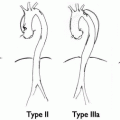
Fig. 25.1
TASC Classification of femoral lesions
5 Diagnosis: Clinical and Laboratory
The diagnosis of femoral artery disease can be made clinically in vast majority of patients. Typical symptoms include intermittent claudication of the calf and limitation of the walking distance. Patients with CFA lesions may also report thigh claudication. Some patients with SFA obstructions and strong profunda collateral describe a “walking-through-phenomenon”: claudication starts after a certain distance at the beginning of the exercise but then disappears during further exercise, and patients are then able to walk pain free for hours.
Occlusion of the SFA in patients with poor collaterals as well as in patients with multi-level disease leads to critical limb ischemia. In patients with single-level disease, even acute occlusion of the SFA frequently causes only an incomplete ischemia, whereas acute occlusion of the entire femoral axis by embolic obstruction of the CFA or both the SFA and DFA always results in symptoms of acute complete ischemia, acutely endangering patient’s limb and life.
Clinical investigation in patients with isolated femoral disease typically reveals a strong pulse at the levels of the groin and a diminished or absent pulse in the popliteal fossa. Auscultation may reveal a bruit of the affected vessel. However, interpretation of vascular bruits remains difficult since harmless plaques particularly in the CFA may cause loud bruits, whereas complete occlusions remain silent.
Despite ongoing scientific efforts, there is still no laboratory test which helps to identify patients with peripheral artery disease. Nevertheless, certain laboratory parameters are helpful when treating these patients:
Traditional risk factors: blood lipids, blood glucose, glycated hemoglobin—to assess the necessity and dosage of adjunctive pharmacological therapy.
Renal function parameters: creatinine—to assess the risk of contrast administration and decide upon renal protective measures before, during, and after an intervention.
Thyroid parameters: TSH—to assess the risk of iodinated contrast administration.
Muscle encymes: creatin kinase and myoglobin—to detect and assess the extent of ischemia of the muscle in patients with acute complete ischemia.
Inflammatory parameters: C-reactive protein, leucocyte count—to identify and assess the extent of inflammation and infection in patients with chronic critical limb ischemia.
Further diagnostic testing should include the following:
(a)
Measurement of the ankle brachial index (ABI) at rest, and optionally ABI after exercise in patients with inconclusive findings at rest.
(b)
Toe pressures have to be measured in patients with diabetic medial sclerosis.
(c)
Oscillography is a useful and quick screening method, which also nicely uncovers the presence of medial sclerosis and can be done in addition to ABI measurements.
6 Diagnostic Imaging
6.1 Duplex Ultrasound Investigation
The femoral segment is the ideal segment for duplex ultrasound investigations. The diagnostic accuracy in skilled hands is about 90%; it is noninvasive and risk free and is an excellent tool to plan endovascular interventions in the entire femoral segments. In our practice, duplex sonography is the only required imaging method for isolated femoral interventions. Furthermore, duplex ultrasound can be used for evaluation of stents or bypasses in the femoral area. Using the peak velocity ratio (PVR: ratio between the maximum systolic flow within the stenosis compared to the systolic velocity in the pre-stenotic segment), an estimation of the degree of stenosis can be given, which nicely corresponds to angiography. Usually, a PVR of 2.4 or 2.5 is considered indicative of a flow-limiting stenosis with a degree of at least 50%. Limitations of duplex ultrasound are as follows:
(a)
Excessive calcifications
(b)
Very obese patients with limited ultrasound visibility
(c)
In patients with proximal flow limitation assessment of the distal vessel can be difficult. Therefore, prior to surgery, advanced imaging methods should be used particularly to assess the distal landing zone of the bypass graft. In the context of bypass surgery, duplex ultrasound also plays an important role in assessing the ipsilateral and contralateral vein situation and suitability as a bypass vessel. A well-trained ultrasound team therefore is mandatory.
6.2 MR Angiography with Gadolinium
(contrast-enhanced magnetic resonance angiography—ce-MRA) can be considered as the new gold standard for evaluation of the lower limb arteries. The diagnostic accuracy particularly in the femoral segment is above 95%, the diagnostic accuracy is not affected by calcification and it can be used to plan endovascular as well as surgical interventions. Nevertheless, there are some limitations of ce-MRA: it tends to overestimate the degree of stenosis—short high-grade stenoses are frequently displayed as complete occlusions. Furthermore, ce-MRA has to be carefully used in patients with severe renal dysfunction due to the risk of nephrogenic fibrosis. In patients with stents, artifacts usually make the interpretation of the remaining lumen problematic. Finally, general contraindications for MR like pacemakers or metal implants have to be considered. In clinical practice, a certain percentage (approximately 5%) of the patients therefore is not suitable for MRA.
6.3 CT Angiography
In contrast to the predominant role of CTA for assessment of aortoiliac disease, heavy calcification frequently limits the usefulness of CTA for assessment of the femoral segment. CTA has the definite advantage of adequately assessing stents and instent-restenosis, but has problems with calcification, includes the use of iodinated contrast administration and radiation.
6.4 Angiography (Digital Subtraction)
This method remains the most accurate way of assessing the vasculature and has to be done routinely immediately before all endovascular interventions. Femoral angiography can be done via an ipsilateral antegrade access by direct puncture of the CFA at the level of the femoral head, by contralateral access via retrograde puncture of the contralateral CFA and an over-the-bifurcation approach or alternatively via a left (preferred) or a right transbrachial or transradial access. Contrast injection can be done manually in doses of 5–10 ml (diluted) contrast, which is the preferred way for selective injection, or via an automated flush injection to the distal aorta, when bilateral overview angiograms are acquired. The usual dosage for an automated overview angiogram to the distal aorta using a pig-tail catheter is 20 ml contrast injected within 10 s.
7 Management
The clinical stage of peripheral artery disease is categorized using the Rutherford–Becker (1–6) or Fontaine (I–IV) classifications. Femoral artery disease should be treated only in symptomatic patients with either intermittent claudication (Rutherford 3) or symptoms of critical limb ischemia (Rutherford 4: ischemic rest pain; Rutherford 5: minor tissue loss; Rutherford 6: major tissue loss). The only exception is patients after peripheral bypass surgery with asymptomatic high-grade stenosis of the bypass or its proximal or distal anastomosis. These patients also have to be treated in an asymptomatic stage to preserve the function of the bypass graft and prevent graft thrombosis; all other patients should be treated only when symptomatic.
Clinical Indications
In patients with intermittent claudication, the indication for treatment is improvement of symptoms, improvement of the walking distance and quality of life. Patients with intermittent claudication have an excellent spontaneous prognosis with respect to limb salvage. Therefore, it has to be clearly stated that in these patients, interventions (or operations) are not necessary to preserve the limb. Patients have to be informed that any intervention at this stage is a “life-style indication.” Nevertheless, considering that intermittent claudication can be a severe burden and may lead to a significant reduction in the quality of life, revascularisation also is justified in patients with severe intermittent claudication. Furthermore, improvement of the walking distance and increase of the exercise capacity also help to improve cardiovascular risk profile.
In patients with critical limb ischemia, revascularisation is mandatory and has to be attempted in almost all patients. Amputation rates in patients with critical limb ischemia with and without successful revascularisation are 5–10% versus 40–75% indicating the huge benefit of revascularisation in these patients. Therefore, in patients with critical limb ischemia, the indication for revascularisation is limb salvage. Exceptions to this rule are patients with permanent paraplegia as well as patients with extensive irreversible tissue necrosis, where primary amputation might be an adequate treatment option.
Indications by Morphology and Location of the Disease
As mentioned above, the CFA traditionally is “surgeon´s territory.” Results of endarterectomy of isolated CFA lesions are clearly better than any endovascular treatment of this segment; therefore, endovascular therapy of the CFA is reserved for selected patients. This includes patients who are unfit for surgery due to general medical conditions, patients with hostile groins (skin infections, very obese patients) as well as patients who are undergoing simultaneous revascularisation procedures of the SFA or DFA. Generally, we try to avoid stenting of the CFA to keep the surgical option. Stenting of the CFA can be done using self-expanding stents and stents later can be even re-punctured for an ipsilateral groin access, but if the stent fails in the CFA a surgical procedure can become very complex.
Indication for endovascular DFA interventions is exclusively given in patients with chronic SFA occlusions to improve the collateral flow when the SFA cannot be reopened.
SFA interventions are done for lesions of any length and grade. Today, the decision for medical treatment, endovascular or surgical treatment will not only depend on the length and morphology of the lesion, but mainly on the clinical stage and patient’s comorbid conditions.
8 List of the Open Operative Choices
Thrombendarterectomy with or without patch of the CFA/DFA/SFA origin
Bypass surgery including femoral-femoral, femoral-popliteal (P1, P3), femoral-crural, femoral-pedal, iliaco-femoral, or ilaco-crural bypass operation. Bypass materials include vein grafts, which have the best patency, or alternatively prosthetic grafts, which are mainly used for supra-genicular anastomosis. Alternatively, hybrid grafts with a prosthetic body and a venous cuff for the distal anastomosis can be used, particularly, for below the knee anastomosis.
Remote endarterectomy with or without endovascular stent support.
9 Intervention: Technique and Pitfalls
9.1 Patient Preparation
Patients receive therapy with aspirin (100 mg) and clopidogrel (75 mg) in preparation for stent implantation. If clopidogrel is started less than 7 days before the intervention, a loading dose with 300 mg is administered 1 day pre-procedure or 600 mg at the day of the procedure. Patients should be well hydrated. During the intervention, we administer 2.000–5.000 IU (rarely 7.000) unfractionated heparin, depending on the length of the intervention. ACT monitoring is not routinely used at our institution.
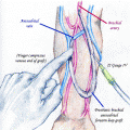
9.2 Access to the Lesion
Generally, we prefer the contralateral crossover approach (Fig. 25.2


Related posts:


Stay updated, free articles. Join our Telegram channel
Full access? Get Clinical Tree
