CHAPTER 22
First-Trimester Fetal Echocardiography
Congenital heart disease is a common condition that accounts for approximately half of perinatal deaths resulting from congenital abnormalities and the majority of neonatal deaths.1 The goal of prenatal diagnosis of congenital heart disease is to optimize prenatal and neonatal care. Preoperative morbidity and mortality rates are lower for prenatally diagnosed cardiac lesions because delivery can be arranged in a center with adequate neonatal resuscitation and surgical expertise.2
The diagnosis of abnormalities of the fetal heart also allows for comprehensive counseling, so parents may make an informed decision about continuing the pregnancy, consider medical therapies, make preparations for potential cardiac surgery, and entertain the possibility of invasive intrauterine intervention if appropriate.3 Last, prenatal diagnosis allows the providers who care for these infants in the delivery room and nurseries to be prepared for problems that might develop after birth.
Fetal echocardiograms performed between 18 and 22 weeks’ gestation remain the gold standard for diagnosis of congenital cardiac anomalies.4 It is during this time frame that the fetus is thought to be large enough to allow a thorough evaluation of the heart but not far enough in gestation to encounter factors that can impede an adequate evaluation. These include acoustic shadowing from fetal bone, fetal position, and decreased amniotic fluid. Eighteen to 22 weeks’ gestation also affords the opportunity to consider in utero intervention or termination when warranted.
Recently, advances in ultrasound equipment functionality and image resolution, and introduction of first-trimester aneuploidy screening, have contributed to an interest in first-trimester fetal echocardiography.5 Earlier fetal echocardiography has known potential benefits, including earlier confirmation of a cardiac abnormality in high-risk patients, allowing for safer termination of pregnancy, longer time frame for fetal karyotyping and genetic counseling, and in some cases earlier initiation of in utero therapy.6
However, first-trimester fetal echocardiography does have limitations and should be considered an adjunct to the second-trimester fetal echocardiogram, not a replacement. The fetal heart is a very small structure at any time in gestation. This is particularly true in the first trimester. Even with the advent of high-resolution transvaginal transducers, identification of all cardiac structures necessary to constitute a complete examination is not possible. Additionally, it is well established that some congenital cardiac lesions, particularly left-sided abnormalities such as hypoplastic left heart syndrome, can be progressive abnormalities, with alterations in myocardial thickening and size of the great vessels occurring later in pregnancy.7 Therefore a normal first-trimester fetal echocardiogram does not exclude congenital heart disease.
Timing
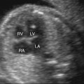
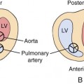
Development of the fetal cardiovascular system occurs by 3 to 6 weeks after conception.1 By 10 weeks’ gestation a four-chambered structure is established.6 Current literature reports considerable variability regarding when fetal cardiac anatomy can be adequately visualized by ultrasonography. Haak et al8 reported cardiac anatomy being adequately evaluated in 20% of cases at 11 weeks’ gestation and 95% of cases at 14 weeks’ gestation. Yagel et al6 reported cardiac anatomy fully discernible in 95% of cases at 11 to 12 weeks’ gestation and in 100% at 13 to 15 weeks’ gestation transvaginally. Lederer et al9 were able to obtain a four-chamber view of the heart in the first trimester in 75 of 130 (57%) cases and outflow tracts in 87 of 130 (59%) cases. Vimpelli et al10 were able to visualize standard views of the fetal heart 58% of the time between 11 weeks and 13 weeks 6 days. Factors influencing visualization include operator experience, equipment capabilities, maternal body habitus, and fetal position.11 Publications reporting experience with early fetal echocardiography suggest that the ideal gestational age to perform the study ranges between 11 and 16 weeks’ gestation.1,5,6,12,13
Accuracy
Yagel et al14 summarized 6000 cases that had a transvaginal fetal echocardiogram at 13 to 16 weeks compared with 15,000 patients scanned only at mid second trimester. The sensitivity of the two approaches was compared. A total of 42 malformations were identified in the group that underwent early echocardiography. Of these, 64% were detected at the first transvaginal scan, with an additional 17% detected in the mid second trimester. Another 4% were detected in the third trimester. Fifteen percent were only diagnosed postnatally. This compared with 80 malformations identified in the group that were only scanned in the mid second trimester. Seventy-eight percent of these abnormalities were diagnosed at the initial echocardiogram and 7% in the third trimester. Six cases were only diagnosed postnatally. In both groups the prenatal detection rate was 85%. The lesions that were missed were thought to be progressive congenital heart defects. This study demonstrates the utility of repeated scans.
Smrcek et al15 reported 2165 cases scanned between 11 and 14 weeks’ gestation. Twenty cardiac anomalies were diagnosed on an early fetal echocardiogram, with an additional nine defects detected in the second trimester and two in the third trimester. This gave an 87% detection rate prenatally.
In contrast, other studies have reported poorer results. Westin et al12 randomized 39,000 women to either a 12-week or 18-week anatomy scan with fetal echocardiography when indicated. They had an 11% detection rate in the 12-week scan group and 15% in the 18-week group.
Delayed or missed diagnoses fall into four categories:
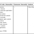
• Inability to visualize the anatomy because of technological limits of resolution. An example of this would be missed diagnosis of ventricular septal defects, which are the most commonly missed lesions on fetal echocardiogram at any gestational age.
• Fetal size and position, which can complicate and compromise the examination, particularly with the limited range of motion of transvaginal transducers.
• Progression of cardiac lesions in utero.
• Operator error. A high level of expertise is necessary to accurately perform any fetal echocardiogram. This may be particularly true when a fetal echocardiogram is attempted in the first trimester.
Intuitively, the more severe the cardiac abnormality, the higher the likelihood it will be diagnosed on first-trimester fetal echocardiography.5
Related posts:
Stay updated, free articles. Join our Telegram channel

Full access? Get Clinical Tree


