Focal gallbladder wall thickening is often an imaging diagnosis and encompasses a wide variety of differential diagnoses. Polypoid lesions of the gallbladder form an important group of conditions that are included in the differential diagnosis of focal gallbladder wall thickening and can be divided into neoplastic and non-neoplastic groups ( Figure 57-1 ). The neoplastic group includes adenomas, leiomyomas, neurofibromas, and gallbladder carcinoma. The non-neoplastic group includes lesions such as cholesterol polyps, inflammatory polyps, adenomyoma, and focal xanthogranulomatous cholecystitis (XGC) ( Box 57-1 ).

Gallbladder Adenoma
Etiology
The majority of gallbladder adenomas are associated with cholelithiasis (50% to 65%). An increased incidence of gallbladder adenomas and biliary tract adenomas are seen in familial adenomatous polyposis and Peutz-Jeghers syndrome.
Prevalence and Epidemiology
Gallbladder adenomas are uncommon lesions, found in 0.5% of cholecystectomy specimens. A small proportion of the gallbladder adenomas can progress to carcinoma, and approximately 10% are multiple.
Clinical Presentation
Gallbladder adenomas are usually asymptomatic and incidentally discovered. Large adenomas or sometimes small adenomas can obstruct the cystic duct and cause right upper quadrant pain.
Pathophysiology
The most common variant is a tubular adenoma. They appear as polypoid structures that project into the gallbladder lumen and may be sessile or pedunculated and generally less than 2 cm.
Imaging
Gallbladder polyps with a stalk and a diameter less than 10 mm are predominantly benign. Sessile polyps and those greater than 10 mm in diameter have a higher likelihood of harboring malignancy and are often an indication for elective cholecystectomy. Adenomas obstructing the cystic duct may lead to gallbladder hydrops or cholecystitis.
Computed Tomography
Gallbladder adenomas are seen as intraluminal soft tissue masses that are isoattenuating or hypoattenuating relative to the liver on contrast-enhanced CT ( Table 57-1 ). These intraluminal masses are difficult to distinguish from noncalcified gallstones on CT, and ultrasonography often helps.
| Modality | Accuracy | Limitations | Pitfalls |
|---|---|---|---|
| Radiography | Data not available to specify accuracy | Insensitive Nonspecific | Unable to directly visualize the soft tissues of gallbladder |
| CT | Data not available to specify accuracy | Radiation exposure Not ideal in pregnant patients | CT may not differentiate noncalcified gallstones from adenomas |
| MRI | Data not available to specify accuracy | Expensive | |
| Ultrasonography | Data not available to specify accuracy | Operator dependent | Differentiation from gallstones adherent to the wall may be difficult |
| Nuclear medicine | Data not available to specify accuracy No role in imaging of adenomas | ||
| PET/CT | Data not available to specify accuracy | Decreased sensitivity in patients with diabetes | Differentiation from gallbladder cancer is not always possible |
Magnetic Resonance Imaging
Polyps are usually seen as homogeneous and low to intermediate signal intensity on T1- and T2-weighted images. Contrast enhancement is seen on delayed images.
Ultrasonography
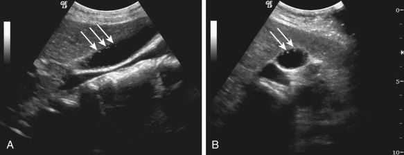
The lesions appear as smoothly marginated, intraluminal polypoid masses with an occasional lobulated or cauliflower-like contour ( Figure 57-2 ). There is a homogenous hyperechoic echotexture, but the echogenicity decreases with increasing size and large adenomas may have a heterogeneous appearance.

The adjacent gallbladder wall characteristically maintains a normal thickness of less than 3 mm. Focal gallbladder wall thickening adjacent to a polypoid mass increases the probability of malignancy.
Gallstones are common in patients with gallbladder adenomas.
Positron Emission Tomography With Computed Tomography
Positron emission tomography (PET) is not usually indicated in the diagnosis of adenomas. However, it has a potential application in ruling out malignancy within a polypoid lesion in the gallbladder.
- •
A smoothly marginated polypoid, sessile, or pedunculated lesion projects into the gallbladder lumen.
- •
Cholelithiasis is a common association.
Differential Diagnosis
These lesions are usually asymptomatic and thus detected incidentally. Adenomas obstructing the cystic duct may present with symptoms of acute cholecystitis (see Chapter 56 ). Gallstones are differentiated based on mobility and adherence to the gallbladder wall. Gallbladder carcinoma has a heterogeneous internal architecture with mucosal irregularity, adjacent parenchymal liver invasion, biliary duct dilatation, metastases, and lymphadenopathy.
Treatment
Because of their malignant potential, cholecystectomy is recommended for gallbladder adenomas larger than 10 mm.
Cholesterol Polyps
Etiology
These benign lesions are uncommonly associated with cholelithiasis and cholesterolosis.
Prevalence and Epidemiology
Cholesterol polyps are benign lesions with no malignant potential that account for approximately 50% of the polypoid lesions in the gallbladder. They predominantly occur in women in their fifth or sixth decades.
Clinical Presentation
Usually asymptomatic, these polyps are typically found in patients who are being evaluated for epigastric discomfort and right upper quadrant pain.
Pathology
The cholesterol polyps are composed of lipid-laden macrophages and are covered by normal gallbladder epithelium that can invaginate and form gland-like structures. They can be single or multiple and usually are less than 10 mm in diameter.
Imaging
Cholesterol polyps are incidental findings on imaging ( Table 57-2 ).
| Modality | Accuracy | Limitations | Pitfalls |
|---|---|---|---|
| Radiography | Data not available to specify accuracy | Insensitive Nonspecific | Unable to directly visualize the soft tissues of gallbladder |
| CT | Data not available to specify accuracy | Radiation exposure Not ideal in pregnant patients | Identification is difficult because the polyps have attenuation similar to bile Differentiation from floating stones and tumefactive sludge can be difficult. |
| MRI | Data not available to specify accuracy | Expensive | |
| Ultrasonography | Data not available to specify accuracy | Operator dependent | |
| Nuclear medicine | Data not available to specify accuracy No role in imaging of adenomas | ||
| PET/CT | Data not available to specify accuracy | Decreased sensitivity in patients with diabetes |
Computed Tomography
On unenhanced scans, cholesterol polyps are often not identifiable because they have attenuation values similar to those of bile. These polyps show enhancement on contrast-enhanced computed tomography (CT) and often appear as floating within the lumen of gallbladder because the thin stalk is not detected on CT and hence are mistaken for noncalcified stones or tumefactive sludge.
Ultrasonography
Small polyps are seen as round or slightly lobulated, brightly echogenic masses attached to a gallbladder wall with no posterior acoustic shadowing ( Figure 57-3 ).
Larger polyps are less echogenic and are characterized by aggregations of echogenic foci within. The presence of these echogenic aggregates within the large polyps is helpful in differentiating cholesterol polyps from benign adenomas and malignant tumors. Large cholesterol polyps can mimic gallbladder carcinoma.
- •
On ultrasonography, cholesterol polyps are seen as single or multiple nodular brightly echogenic masses attached to the gallbladder wall.
- •
Posterior acoustic shadowing is not present.
- •
Cholesterol polyps show enhancement on contrast-enhanced CT and are seen as floating within the gallbladder lumen.
Differential Diagnosis
Cholesterol polyps are usually asymptomatic. Gallstones adherent to the gallbladder wall are echogenic and show posterior acoustic shadowing. These are often mistaken for cholesterol polyps. Tumefactive sludge forms another differential diagnosis but can be discerned by the difference in morphology as shown with a change in position of the patient.
Adenomas are smooth, lobulated or rounded masses with a homogeneous echotexture and an identifiable stalk in pedunculated polyps. Gallbladder carcinoma is evident by its heterogeneous internal architecture with mucosal irregularity, adjacent parenchymal liver invasion, biliary duct dilatation, metastases, and lymphadenopathy.
Treatment
Small cholesterol polyps are conservatively treated with follow-up. Large polyps mimicking gallbladder carcinoma may require cholecystectomy to rule out malignancy.
Cholesterol Polyps
Etiology
These benign lesions are uncommonly associated with cholelithiasis and cholesterolosis.
Prevalence and Epidemiology
Cholesterol polyps are benign lesions with no malignant potential that account for approximately 50% of the polypoid lesions in the gallbladder. They predominantly occur in women in their fifth or sixth decades.
Clinical Presentation
Usually asymptomatic, these polyps are typically found in patients who are being evaluated for epigastric discomfort and right upper quadrant pain.
Pathology
The cholesterol polyps are composed of lipid-laden macrophages and are covered by normal gallbladder epithelium that can invaginate and form gland-like structures. They can be single or multiple and usually are less than 10 mm in diameter.
Imaging
Cholesterol polyps are incidental findings on imaging ( Table 57-2 ).
| Modality | Accuracy | Limitations | Pitfalls |
|---|---|---|---|
| Radiography | Data not available to specify accuracy | Insensitive Nonspecific | Unable to directly visualize the soft tissues of gallbladder |
| CT | Data not available to specify accuracy | Radiation exposure Not ideal in pregnant patients | Identification is difficult because the polyps have attenuation similar to bile Differentiation from floating stones and tumefactive sludge can be difficult. |
| MRI | Data not available to specify accuracy | Expensive | |
| Ultrasonography | Data not available to specify accuracy | Operator dependent | |
| Nuclear medicine | Data not available to specify accuracy No role in imaging of adenomas | ||
| PET/CT | Data not available to specify accuracy | Decreased sensitivity in patients with diabetes |
Related posts:


Stay updated, free articles. Join our Telegram channel

Full access? Get Clinical Tree


