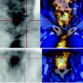
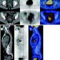
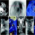
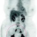
Fig. 77.1
The solid lung mass in the right postero-basal segment, which was already examined with a FNAB, shows pathological increase in the consumption of glucose, SUV max 11.5
No areas of abnormal metabolism in other parts of the body examined.
77.4 Conclusions
The PET scan confirms the presence of a right lung mass characterized by high metabolism, compatible with NSCLC, however, to be studied histologically. See Figs. 77.2, 77.3.
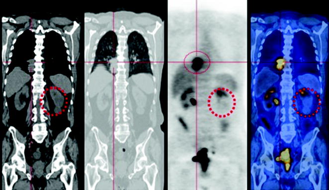

Fig. 77.2


The PET-CT scan shows a heterogeneous mass in the posterior basal segment of the right lung that invades surrounding tissues and appears inseparable from the hilum. The metabolism of glucose of the lesion is high. At the CT scan it is evident a cystic dysplasia of the left lower renal district, characterized by reduced parenchymal function
Stay updated, free articles. Join our Telegram channel

Full access? Get Clinical Tree


