Foot Ultrasound
KEY FACTS
GENERAL CONSIDERATIONS
TECHNIQUE
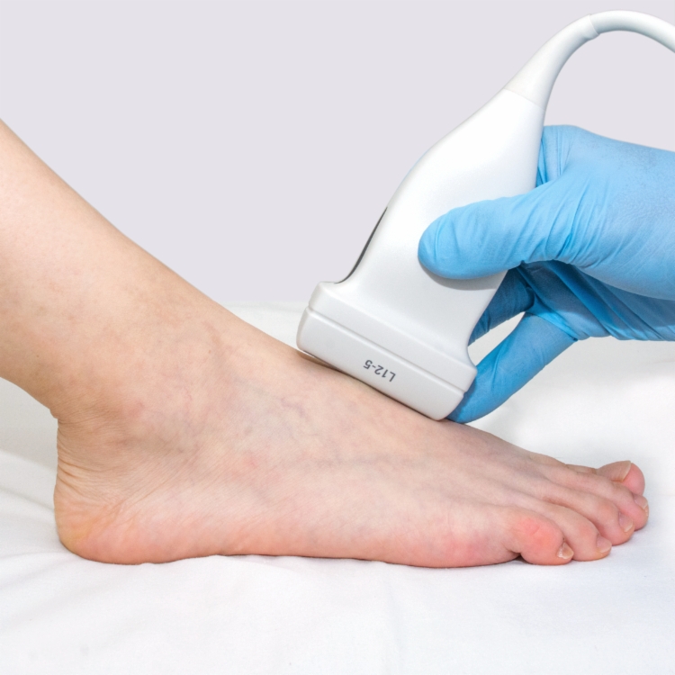
Clinical photograph shows the probe and patient position for the dorsum of the foot.

Longitudinal US shows the bones of the forefoot and midfoot with the talus  , navicular
  , and medial cuneiform
  . The location and shape of the foot bones makes them quite readily recognizable.

Longitudinal US in medial aspect of tarsus shows the base of the 1st metatarsal  , medial cuneiform
  , and navicular
  bones.

Transverse US of the diverging extensor digitorum tendon slips  overlying the extensor hallucis brevis
  and extensor digitorum brevis
  muscles is shown.
GENERAL CONSIDERATIONS
Clinical Indications for Foot US
TECHNIQUE: DORSAL FOOT
Patient Position
Specifically Examine Bones and Joints
 Evaluated from both dorsal and plantar aspects, primarily in longitudinal plane
 Passive flexion and extension can help in assessment
 On plantar aspect, assess plantar plate, paired sesamoid bones, and intervening flexor hallucis longus (FHL)


