Foreign Body and Injection Granuloma
KEY FACTS
Terminology
Imaging
 Reflectivity of some objects (wood, metal, glass, plastic) exceeds that of others (bamboo)
 Align probe as parallel to FB as possible; FB may be located some distance from entry wound
 Echogenic FB ± posterior artifact (reverberation/comet-tail artifact or posterior shadowing)
 ± hypoechoic rim of edematous inflammatory tissue, granulation tissue, or fibrosis
 False-positive US with gas or calcification in soft tissues; correlate with radiographs if necessary
 Repeat US in 2-10 days if clinical history is suspicious and US findings negative or equivocal
IMAGING
General Features
Ultrasonographic Findings
Imaging Recommendations
 FB
FB
![]()
Stay updated, free articles. Join our Telegram channel

Full access? Get Clinical Tree


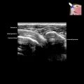
 in the subcutaneous tissues of the hand 7 days after a penetrating skin injury. A rim of hypoechoic granulation tissue
  surrounds the wood splinter.
 in the adjacent granulation tissue and subcutaneous tissues. This foreign body (FB) was successfully removed under US guidance.
 within the edematous thenar muscle
  1 day after a penetrating injury.
 . The thenar musculature
  is also less hypoechoic than previously. Follow-up US is particularly useful for suspected organic FBs not visible on initial US examination.