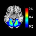
div style=”display:none;”> Frontal Pole (Area 10) Main Text TERMINOLOGY Abbreviations • Frontal pole (Fp) Location and Boundaries Location • Most rostral portion of prefrontal cortex • Includes frontomarginal sulcus, rostral part of superior frontal gyrus, and small parts of middle frontal gyrus Boundaries • No precise anatomic landmarks are available Bounded rostrally and laterally by middle frontal area (area 46) and superior frontal gyrus (area 9) Bounded caudally by area 32 rostral to cingulate gyrus Bounded ventrally by area 11 (rostral end of olfactory sulcus) • 2 distinct cytoarchitectonic regions are present in area 10 Fp1 extends further laterally and rostrally Fp2 occupies medial Fp and is more caudal than Fp1 Function Fp1 • Cognition • Working memory • Perception Fp2 • Affective processing • Mentalizing • Social cognition Frontal Pole Function • Electrophysiology in macaques suggests monitoring of action outcomes • Lesion data Fp involved in goal-directed behaviors and making future choices • Proposed function Maintenance of goals during their deferral while individual engages in multitasking Comparative Anatomy • Expanded size and cell density in humans compared to nonhuman primates • Phylogenetically young region Abstraction • More abstract cognitive thought is performed more anteriorly within prefrontal cortex with frontopolar cortex at apex Structural Connections Tracer/Diffusion Tensor Imaging Studies • Poor characterization of distant area 10 inputs/outputs in human and macaque • Reciprocal connections are with dorsolateral, orbitofrontal, and medial prefrontal cortex • Hierarchical, rostrocaudal connectivity Rostral frontal areas connected to intermediate ones that, in turn, connect to more caudal frontal areas Functional Connections Coactive Regions • Medial Fp (especially Fp2) is coactive with default mode network Posterior cingulate cortex (area 23, 31) Inferior parietal lobule (areas 39, 40) Inferior temporal gyrus (area 20) Associated Literature Keywords (NeuroSynth) • Error related, intentions, remember, thoughts, contextual, salient, autobiographical, amount, correct, stress, nonverbal Area 10-Associated Disorders Anterior Cerebral Artery Ischemia • Common site of injury along with other medial prefrontal and cingulate regions (especially frontopolar branch) Traumatic Injury • Damage to Fp does not impair performance in well-learned, cognitively demanding tasks • Can cause behavioral changes for situations requiring redistribution of resources to novel tasks Image Gallery Print Images BRODMANN AREA 10: FRONTAL POLE Coactivation map of Brodmann area 10 shows brain regions that reliably activate with the centroid of voxels lying within area 10 in over 4,000 studies from the NeuroSynth database. This image shows the average of left and right coactivation maps. Only gold members can continue reading. Log In or Register to continue Share this:Click to share on Twitter (Opens in new window)Click to share on Facebook (Opens in new window) Related posts: Intracranial Arteries Overview Glossopharyngeal Nerve (CNIX) Retrosplenial Cingulate Cortex (Areas 29, 30) Hypoglossal Nerve (CNXII) Medulla Ventricles and Choroid Plexus Stay updated, free articles. Join our Telegram channel Join Tags: Imaging Anatomy Brain and Spine Mar 13, 2021 | Posted by admin in NUCLEAR MEDICINE | Comments Off on Frontal Pole (Area 10) Full access? Get Clinical Tree