KEY FACTS
Imaging
- •
US for initial detection and characterization, CECT or MR for preoperative assessment and staging
- •
3 main morphological types
- ○
Large soft tissue mass infiltrating gallbladder (GB) fossa/replacing GB, ± invading liver ( most common )
- ○
Diffuse or focal GB wall thickening: Asymmetric, irregular, extensive thickening
- ○
Polypoid intraluminal mass: > 1 cm, thickened base, irregular margins
- ○
Top Differential Diagnoses
- •
GB polyp
- •
Chronic or xanthogranulomatous cholecystitis
- •
Hyperplastic cholecystoses
- •
Diffuse GB thickening from portal hypertension, heart failure
Pathology
- •
Most are adenocarcinoma; mean 5-year survival: 5-10%
- •
Chronic irritation of GB mucosa by gallstones (GS)
- •
Malignant degeneration of adenomatous polyps less common
- •
Spreads by local invasion or hematogenous spread to liver, nodal spread to porta hepatis and paraaortic nodes
Clinical Issues
- •
Most common malignancy of biliary tree; prevalence: 3-7%
- •
F:M = 3:1; mean age: 65 years
- •
Risk factors: GS, chronic infection and inflammation
- •
Preoperative diagnosis occurs in < 20% of patients, found at cholecystectomy for stones/cholecystitis
- •
Right upper quadrant pain, weight loss, anorexia, fever
- •
Jaundice occurs when tumor invades bile ducts
Scanning Tips
- •
Look for asymmetric mass infiltrating liver
 : Direct tumor infiltration to liver parenchyma
  and retrograde spread along biliary tree
  .
.
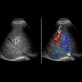
 filled with solid tumor proven to be adenocarcinoma. Color Doppler would be useful to confirm that this is a tumor and not sludge.
 surrounded by tumor
  . The interface
  between the gallbladder and the liver is indistinct, indicating local invasion.
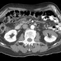
 and kidney
  in the same patient shows liver metastases
  .
.
Stay updated, free articles. Join our Telegram channel

Full access? Get Clinical Tree


