KEY FACTS
Imaging
- •
Single or multiple small, round/ovoid masses attached to gallbladder (GB) wall with no posterior acoustic shadowing
- ○
Usually sessile but may be pedunculated with well-defined stalk
- ○
- •
Usually 2-10 mm in size, most commonly in middle 1/3 of GB
- •
Cholesterol polyp: Small with comet-tail artifact
- •
Avascular or hypovascular on Doppler examination
- ○
Larger lesions may have slight internal vascularity
- ○
- •
Consider neoplastic GB polyp if size > 10 mm, irregular outline, sessile morphology with abnormal GB wall and invasion of adjacent structures, growth on serial US examinations
Top Differential Diagnoses
- •
Hyperplastic cholecystosis/adenomyomatosis
- •
Nonshadowing cholelithiasis or sludge
- •
Adenoma
- •
Carcinoma or metastasis
Pathology
- •
Polypoid lesions include cholesterol polyp, adenomyomatosis, and neoplasms such as adenoma, carcinoma, and metastases
Clinical Issues
- •
5% of population have polyps; 50% are cholesterol polyps
- •
More common in middle age; F > M; incidental finding
- ○
< 6 mm: No follow-up
- ○
7-9 mm: Yearly US follow-up to monitor size
- ○
> 10 mm: Surgical consult
- ○
Scanning Tips
- •
Scan in supine, decubitus (left or right lateral) positions to demonstrate immobility of GB polyp
- •
Look for color flow in polyp and for any abnormality of adjacent GB wall, which might indicate cancer
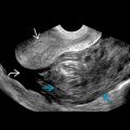
 arising from the gallbladder (GB) wall, compatible with cholesterol polyps. Note the preserved GB wall without invasion to the adjacent liver parenchyma.
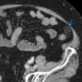
 . There are also dependent stones
  with acoustic shadowing.
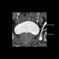
 . This was an incidental finding in a patient with chronic liver disease and ascites
  .
.
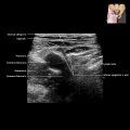
 in the GB neck. Given its size, follow-up is recommended.
Related posts:


Stay updated, free articles. Join our Telegram channel

Full access? Get Clinical Tree


