KEY FACTS
Imaging
- •
Germinal matrix hemorrhage (GMH) most common at caudothalamic (CT) groove in vascular germinal matrix
- •
Grading system
- ○
Grade 1 : GMH only with globular echogenic focus in CT groove
- ○
Grade 2 : GMH + intraventricular hemorrhage (IVH), normal ventricle size
- –
Look for fluid-debris levels in dependent occipital horns
- –
- ○
Grade 3 : GMH + IVH + ventricular expansion
- –
Ventricular expansion some days after grade 2 IVH = secondary hydrocephalus
- –
- ○
Grade 4 : GMH + IVH + intraparenchymal hemorrhage
- –
Venous compression leads to periventricular hemorrhagic venous infarction
- –
Fan-shaped, echogenic, can liquefy, infarcted brain may be replaced by porencephalic cyst
- –
- ○
Top Differential Diagnoses
- •
Hypoxic-ischemic encephalopathy: Ischemic injury due to perinatal insult
- ○
Abnormal echogenicity of affected deep gray &/or white matter
- ○
Heals as periventricular leukomalacia with Swiss cheese pattern in deep white matter
- ○
Scanning Tips
- •
Use small-footprint, high-frequency transducers with multiple focal zones
- •
Use multiple points of access (anterior/posterior fontanel, mastoid, temporal bone)
- ○
Look for fluid levels, blood in 3rd, 4th ventricles, cerebellar hemorrhage
- ○
Any grade of GMH has worse prognosis in setting of coexistent cerebellar hemorrhage
- ○
- •
Cine clips, color Doppler help differentiate avascular, variable-echogenicity clot from echogenic choroid
 are comma-shaped, on either side of the cavum septi pellucidi
  , and connected to the 3rd ventricle by the foramina of Monro
  . The corpus callosum
  is well seen.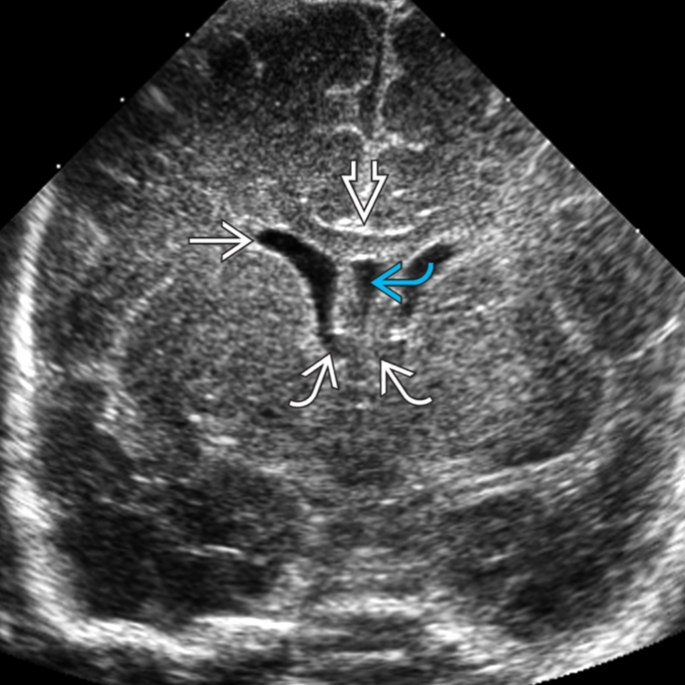
 (caudate
  , thalamus
  ). Normal, echogenic choroid
  never extends anterior to the caudothalamic groove or into the occipital horn.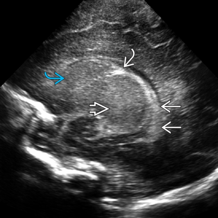
 . This is a grade 1 germinal matrix hemorrhage (GMH).
Related posts:

Stay updated, free articles. Join our Telegram channel

Full access? Get Clinical Tree


