Gout and Pseudogout
Get Clinical Tree app for offline access
IMAGING
General Features
Imaging Recommendations

Gout and Pseudogout
Get Clinical Tree app for offline access
IMAGING
General Features
Imaging Recommendations


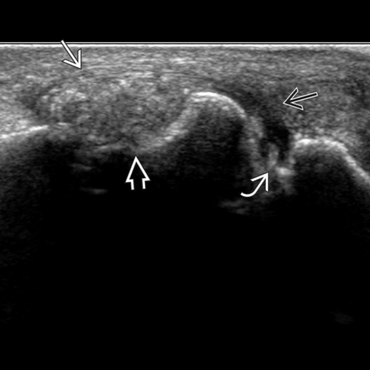
 causes marginal erosion
  of the metatarsal head. A small joint effusion
  is present in the MTPJ with echogenic gouty crystal aggregates
  .
.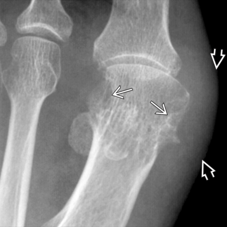
 and marginal erosions
  . The tophi are largely noncalcified and not clearly seen.
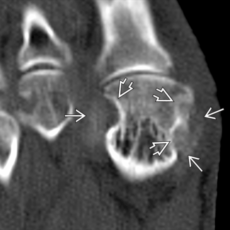
 . CT is more sensitive than radiography at revealing gouty tophi. There are marginal erosions
  deep to the tophi.
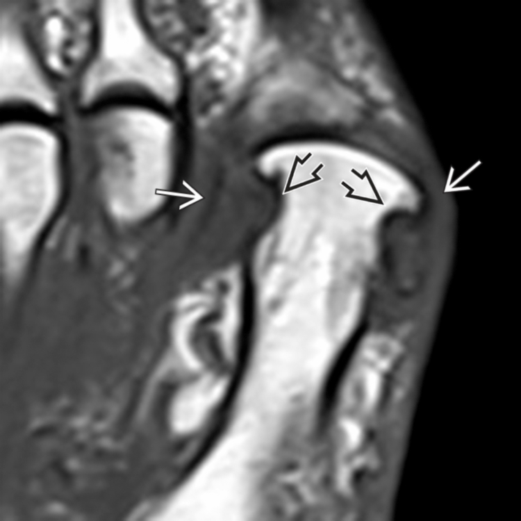
 and marginal erosions
  . Dual-energy CT (if available) is better than MR or US at quantifying gouty tophi volume for monitoring response to treatment.