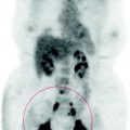
Fig. 40.1
No areas of abnormal metabolism
40.4 Conclusions
The scan shows mediastinal lymphadenopathy, probably of granulomatous nature.
Histological exam is necessary to define the treatment program and completely exclude a neoplastic condition.
Mediastinoscopy was then performed and allowed the removal of two lymph nodes, on which a nonspecific granulomatous process was demonstrated.
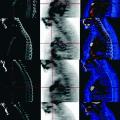
The CT shows some mediastinal lymph node size of increased dimension (Fig. 40.2), in the pre-tracheal (Fig. 40.2a, blue arrows), the aortopulmonary window (Fig. 40.2a, yellow arrows) and in the prevascular area (Fig. 40.2a, b, red arrows). See also Figs. 40.3, 40.4.
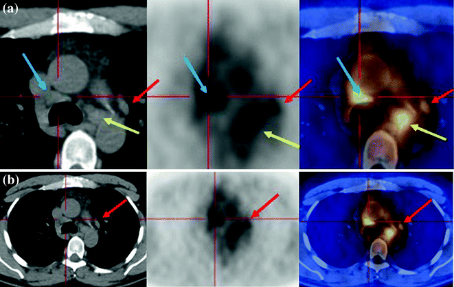
Fig. 40.2
 Laryngeal Squamous Carcinoma: Staging
 Radio-Treated Cancer of the Posterior Hemi-Circumference of the Anal Canal: Post-Actinic Fibrosis
 Bone-Destroying Metastases in Thyroid Undifferentiated Carcinoma
 Surgically Treated Endometrial Cancer: Bilateral Nodal Recurrence
 Undifferentiated Gastric Adenocarcinoma with Peritoneal Carcinosis
 Bone Metastases from Breast Cancer: Progression of Disease and Subsequent Response to Radiotherapy


The CT shows some mediastinal lymph node size of increased dimension in the pre-tracheal (a, blue arrows), the aorto-pulmonary window (a, yellow arrows) and in the prevascular area (a, b, red arrows)
Related posts:
Laryngeal Squamous Carcinoma: Staging
Radio-Treated Cancer of the Posterior Hemi-Circumference of the Anal Canal: Post-Actinic Fibrosis
Bone-Destroying Metastases in Thyroid Undifferentiated Carcinoma
Surgically Treated Endometrial Cancer: Bilateral Nodal Recurrence
Undifferentiated Gastric Adenocarcinoma with Peritoneal Carcinosis
Bone Metastases from Breast Cancer: Progression of Disease and Subsequent Response to Radiotherapy
Stay updated, free articles. Join our Telegram channel
Full access? Get Clinical Tree
