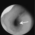
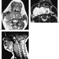
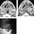
Chapter 205 The group (level) I lymph nodes are situated above the hyoid bone, superficial to the mylohyoid muscles and anterior to a transverse line drawn on each axial image through the posterior edge of the submandibular gland (Fig. 205–1). Nodes are considered level I if the majority of the cross-sectional area of the node lies anterior to a line drawn tangential along the posterior edge of the submandibular gland. A node is considered level II if the majority of the cross-sectional area of the gland is located posterior to this line. Level I nodes can be subclassified into levels IA and IB. Level IA (submental) are situated between the medial margins of the anterior bellies of the digastric muscle, below the mylohyoid muscle and above the hyoid bone. Level IB (submandibular) are located posterior and lateral to the medial edge of the anterior belly of the digastric muscle, below the mylohyoid muscle, above the hyoid bone, and anterior to a transverse line drawn tangential to the posterior surface of the submandibular gland. The afferent drainage is from the oral cavity, sinonasal cavity, and facial nodes. Normal level I nodes can be routinely seen on imaging. Level I nodes are longitudinally oriented, whereas levels II through V are vertically oriented. Thus, it is possible to visualize an eccentric fatty hilum on transverse imaging. This should not be confused with a peripheral metastasis or an area of necrosis. Level I nodes can be enlarged by a variety of pathological processes (Figs. 205–2 and 205–3
Group (Level) I Lymph Nodes (Submandibular–Submental)
Epidemiology
Clinical Features
Pathology
![]()
Stay updated, free articles. Join our Telegram channel


Full access? Get Clinical Tree


