The Skull and Facial Bones
The skull consists of the calvarium, facial bones and mandible. The calvarium is the brain case and comprises the skull vault and skull base. The bones of the calvarium and face are joined at immovable fibrous joints, except for the temporomandibular joint, which is a movable cartilaginous joint.
THE SKULL VAULT ( Figs. 1.1–1.4 )
The skull vault is made up of several flat bones, joined at sutures, which can be recognized on skull radiographs. The bones consist of the diploic space – a cancellous layer containing vascular spaces – sandwiched between the inner and outer tables of cortical bone. The skull is covered by periosteum, which is continuous with the fibrous tissue in the sutures. The periosteum is called the pericranium externally and on the deep surface of the skull it is called the endosteum. The endosteum is the outer layer of the dura. The diploic veins within the skull are large, valveless vessels with thin walls. They communicate with the meningeal veins, the dural sinuses and the scalp veins.


- 1.
Coronal suture
- 2.
Zygomaticofrontal suture
- 3.
Pterion
- 4.
Sphenotemporal (sphenosquamosal) suture
- 5.
Temporoparietal (squamosal) suture
- 6.
Asterion
- 7.
Lambdoid suture
- 8.
Wormian bones
- 9.
Lambda
- 10.
Sagittal suture
- 11.
Anterior fontanelle
- 12.
Metopic suture
- 13.
Nasofrontal suture
- 14.
Zygomaticofrontal suture

- 1.
Bregma
- 2.
Coronal suture
- 3.
Lambda
- 4.
Lambdoid suture
- 5.
Vertex
- 6.
Inner skull table
- 7.
Outer skull table
- 8.
Internal occipital protuberance
- 9.
External occipital protuberance
- 10.
External auditory meatus
- 11.
Styloid process
- 12.
Clivus
- 13.
Dorsum sellae
- 14.
Posterior clinoid process
- 15.
Anterior clinoid process
- 16.
Pituitary fossa (sella turcica)
- 17.
Tuberculum sellae
- 18.
Planum sphenoidale
- 19.
Greater wings of sphenoid
- 20.
Undulating floor of anterior cranial fossa (roof of orbit)
- 21.
Anterior limit of foramen magnum
- 22.
Posterior limit of foramen magnum
- 23.
Posterior wall of maxillary sinus
- 24.
Floor of orbit
- 25.
Hard palate
- 26.
Neck of mandible
- 27.
Temporomandibular joint
- 28.
Condylar (mandibular) canal
Vascular markings
- 29.
Middle meningeal vessels: anterior branches
- 30.
Middle meningeal vessels: posterior branches
- 31.
Transverse sinus
- 32.
Diploic vein
- 33.
Diploic venous confl uence: parietal star
Sinuses/air cells
- 34.
Frontal sinus
- 35.
Sphenoid sinus
- 36.
Posterior ethmoidal cells
- 37.
Maxillary sinus
- 38.
Mastoid air cells
Soft tissues
- 39.
Soft palate
- 40.
Base of tongue

- 1.
Sagittal suture
- 2.
Frontal sinus
- 3.
Planum sphenoidale
- 4.
Crista galli
- 5.
Perpendicular plate of ethmoid
- 6.
Floor of pituitary fossa
- 7.
Nasal septum
- 8.
Ethmoid air cells
- 9.
Superior orbital fissure
- 10.
Lesser wing of sphenoid
- 11.
Innominate line
- 12.
Zygomatic process of frontal bone
- 13.
Zygomaticofrontal suture
- 14.
Frontal process of zygomatic bone
- 15.
Foramen rotundum
- 16.
Petrous ridge
- 17.
Maxillary sinus
- 18.
Inferior nasal turbinate
- 19.
Mastoid process
- 20.
Occipital bone
- 21.
Dens of atlas
The paired parietal bones form much of the side and the roof of the skull and are joined in the midline at the sagittal suture. Parietal foramina are paired foramina or areas of thin bone close to the midline in the parietal bones. They are often visible on a radiograph, may be big and may even be palpable. They may transmit emissary veins from the sagittal sinus. The frontal bone forms the front of the skull vault. It is formed by two frontal bones that unite at the metopic suture. The frontal bones join the parietal bones at the coronal suture. The junction of coronal and sagittal sutures is known as the bregma . The occipital bone forms the back of the skull vault and is joined to the parietal bones at the lambdoid suture. The lambdoid and sagittal sutures join at a point known as the lambda .
The greater wing of sphenoid and the squamous part of the temporal bone form the side of the skull vault below the frontal and parietal bones. The sutures formed here are: (1) the sphenosquamosal suture between the sphenoid and temporal bones; (2) the sphenofrontal and sphenoparietal sutures between the greater wing of the sphenoid and frontal and parietal bones; and (3) the squamosal suture between the temporal and parietal bones. The sphenofrontal, sphenoparietal and squamosal sutures form a continuous curved line (see Fig. 1.1 ). The intersection of the sutures between the frontal, sphenoidal, parietal and temporal bones is termed the pterion and provides a surface marking for the anterior branch of the middle meningeal artery on the lateral skull radiograph. The asterion is the point where the squamosal suture meets the lambdoid suture.
The parietal and occipital bones develop from multiple ossification centres and may have accessory sutures , particularly in childhood, that can persist into adulthood. An accessory intraparietal suture may extend superolaterally from the lambdoid suture into the parietal bone. Mendosal sutures in the occipital bone extend for a variable distance superomedially from the inferior part of the lambdoid sutures. An intraoccipital accessory suture may be seen crossing horizontally just above the foramen magnum.
Accessory sutures, more common in children, should not be mistaken for skull fractures. Accessory sutures, compared with fractures, can be seen on computed tomography (CT) to have a zig-zag configuration with sclerotic borders, to arise from but not cross suture lines, and are often bilateral and symmetrical.
THE SKULL BASE ( Figs. 1.5, 1.6 )
The inner aspect of the skull base is made up of the following bones from anterior to posterior:
- ■
The orbital plates of the frontal bone, with the cribriform plate of the ethmoid bone and crista galli in the midline
- ■
The sphenoid bone with its lesser wings anteriorly forming the posterior part of the floor of the anterior cranial fossa, the greater wings posteriorly forming the anterior part of the floor of the middle cranial fossa. The body of the sphenoid bone with the elevated sella turcica is in the midline
- ■
Part of the squamous temporal bone and the petrous temporal bone, and
- ■
The occipital bone

- 1.
Crista galli
- 2.
Anterior clinoid process
- 3.
Optic canal
- 4.
Posterior clinoid process
- 5.
Cribriform plate
- 6.
Posterior ethmoidal foramen
- 7.
Foramen ovale
- 8.
Foramen spinosum
- 9.
Foramen lacerum
- 10.
Jugular foramen
- 11.
Foramen magnum
- 1.
Greater palatine foramen
- 2.
Pterygoid plate
- 3.
Foramen ovale
- 4.
Foramen spinosum
- 5.
External acoustic foramen
- 6.
Jugular fossa
- 7.
Foramen lacerum
- 8.
Groove for pharyngotympanic tube
- 9.
Styloid process
- 10.
Stylomastoid foramen
- 11.
Foramen magnum

- 1.
Right internal auditory meatus
- 2.
Right jugular foramen
- 3.
Right carotid canal
- 4.
Right foramen ovale
- 5.
Right foramen spinosum
- 6.
Right pterygopalatine fossa
- 7.
Right hypoglossal canal
INDIVIDUAL BONES OF THE SKULL BASE
The orbital plates of the frontal bones are thin and irregular, and separate the anterior cranial fossa from the orbital cavity.
The cribriform plate of the ethmoid bone is a thin, depressed bone separating the anterior cranial fossa from the nasal cavity. It has a superior perpendicular projection, the crista galli, which is continuous below with the nasal septum on the frontal skull radiograph (see Fig. 1.4 ).
The sphenoid bone consists of a body and greater and lesser wings, which curve laterally from the body and join at the sharply posteriorly angulated sphenoid ridge. The body houses the sphenoid sinuses and is grooved laterally by the carotid sulcus, in which the cavernous sinus and carotid artery run. The sphenoid body has a deep fossa superiorly ( Figs. 1.7 and 2.14) known as the sella turcica or pituitary fossa , which houses the pituitary gland. On the anterior part of the sella is a prominence known as the tuberculum sellae ; anterior to this is a groove called the sulcus chiasmaticus , which leads to the optic canal on each side. The optic chiasm lies over this sulcus. Two bony projections on either side of the front of the sella are called the anterior clinoid processes . The posterior part of the sella is called the dorsum sellae , and this is continuous posteriorly with the clivus . Two posterior projections of the dorsum sellae form the posterior clinoid processes . The floor of the sella is formed by a thin bone known as the lamina dura , which may be eroded by raised intracranial pressure or tumours of the pituitary.

The temporal bone consists of four parts:
- ■
A flat squamous part, which forms part of the vault and part of the skull base
- ■
A pyramidal petrous part, which houses the middle and inner ears and forms part the boundary between the middle and posterior cranial fossae
- ■
An aerated mastoid part, and
- ■
An inferior projection known as the styloid process
The zygomatic process projects from the outer side of the squamous temporal bone and is continuous with the zygomatic process of the zygoma to form the zygomatic arch.
The curved occipital bone forms part of the skull vault and posterior part of the skull base. It has the foramen magnum in the midline, through which the cranial cavity is continuous with the spinal canal. Anterolaterally it is continuous with the posterior part of the petrous bones on each side, and anterior to the foramen magnum it forms the clivus. The clivus is continuous anteriorly with the dorsum sellae. Thus the occipital bone articulates with both the temporal and sphenoid bones. The occipital condyles for articulation with the atlas vertebra project from the inferior surface of the occipital bone lateral to the anterior half of the foramen magnum.
CRANIAL FOSSAE (see Fig. 1.5 )
The anterior cranial fossa is limited posteriorly by the sphenoid ridge and anterior clinoid processes, and supports the frontal lobes of the brain.
The middle cranial fossa is limited anteriorly by the sphenoid ridge and anterior clinoid processes. Its posterior boundary is formed laterally by the petrous ridges and in the midline by the posterior clinoid processes and dorsum sellae. It contains the temporal lobes of the brain, the pituitary gland and most of the foramina of the skull base.
The posterior cranial fossa is the largest and deepest fossa. Anteriorly it is limited by the dorsum sellae and the petrous ridge, and it is demarcated posteriorly on the skull radiograph by the groove for the transverse sinus. It contains the cerebellum posteriorly, and anteriorly the pons and medulla lie on the clivus and are continuous, through the foramen magnum, with the spinal cord.
FORAMINA OF THE SKULL BASE (see Figs. 1.5, 1.6 ; Table 1.1 )
The optic canals run from the sulcus chiasmaticus anterior to the tuberculum sellae, anteroinferolaterally to the orbital apex. They transmit the optic nerves and ophthalmic arteries. The optic canals are wider posteriorly than anteriorly.
The close proximity of the optic canal to the sphenoid sinus is important in planning paranasal sinus surgery.
| Comment | Transmits | ||
|---|---|---|---|
| Optic canals | Sphenoid bone | Middle cranial fossa to orbital apex | Optic nerves and ophthalmic arteries |
| Superior orbital fissure | Sphenoid bone | From the middle cranial fossa to the orbital apex | First (orbital) division of the fifth, and the third, fourth and sixth cranial nerves, superior orbital vein and a branch of the middle meningeal artery |
| Inferior orbital fissure | Between maxilla and sphenoid bones | At its posterior part it forms an opening between the orbit and the pterygopalatine fossa, and more anteriorly between the orbital cavity and the infratemporal fossa | Infraorbital nerve and infraorbital artery and the inferior ophthalmic veins |
| Foramen rotundum | Sphenoid bone | From the middle cranial fossa to the pterygopalatine fossa | Second (maxillary) division of the fifth cranial nerve |
| Foramen ovale | Sphenoid bone | From the middle cranial fossa to the infratemporal fossa | Third (mandibular) division of the fifth cranial nerve and the accessory meningeal artery |
| Foramen spinosum | Sphenoid bone | From the infratemporal to the middle cranial fossa | The middle meningeal artery |
| Foramen lacerum | Apex of temporal bone | Carotid artery passes through its posterior wall | |
| IAM | Petrous temporal bone | From the posterior cranial fossa to inner ear | Seventh and eighth cranial nerves and the internal auditory artery |
| Jugular foramen | Junction of occipital and petrous temporal bones | Not visible on SMV | Internal jugular vein, ninth, tenth and eleventh cranial nerves. Inferior petrosal sinus (which drains into the internal jugular vein), and ascending occipital and pharyngeal arterial branches |
| Hypoglossal canal | Occipital bone | 12th (hypoglossal) cranial nerve | |
| Foramen magnum | Occipital bone | From the posterior cranial fossa to the spinal canal | Medulla oblongata/spinal cord, along vertebral and spinal arteries and veins, and the spinal root of the 11th cranial nerve |
The superior orbital fissure is a triangular defect between the greater and lesser wings of the sphenoid. It transmits the first (orbital) division of the fifth cranial nerve, and the third, fourth and sixth cranial nerves, along with the superior orbital vein and a branch of the middle meningeal artery from the middle cranial fossa to the orbital apex. This fissure is best seen on the OF20 view (occipitofrontal view with 20 degrees caudal angulation) and on CTs of the orbit (see Fig. 1.4 and later Fig. 1.10 ).
The foramen rotundum is posterior to the superior orbital fissure in the greater wing of the sphenoid. It runs from the middle cranial fossa to the pterygopalatine fossa and transmits the second (maxillary) division of the fifth cranial nerve. It is seen on the OF20 view (see Fig. 1.4 ) and also on CT scans (see Fig. 1.6 ). See also ‘The infratemporal space and the pterygopalatine fossa’ section (see later Fig. 1.36 ) for this and sphenopalatine foramen and greater palatine foramen .
The foramen ovale is posterolateral to the foramen rotundum in the greater wing of the sphenoid. It runs from the middle cranial fossa to the infratemporal fossa and transmits the third (mandibular) division of the fifth cranial nerve and the accessory meningeal artery.
The foramen spinosum , posterolateral to the foramen rotundum, is a small foramen and transmits the middle meningeal artery from the infratemporal to the middle cranial fossa.
The foramen lacerum is a ragged bony canal posteromedial to the foramen ovale at the apex of the petrous bone. The internal carotid artery passes through its posterior wall, having emerged from the carotid canal (which runs in the petrous bone), before turning upwards to run in the carotid sulcus.
The internal auditory meatus (IAM) and canal run from the posterior cranial fossa through the posterior wall of the petrous bone into the inner ear. They transmit the seventh and eighth cranial nerves and the internal auditory artery. The jugular foramen is an irregular opening situated at the posterior end of the junction of the occipital and petrous bones. It runs downward and medially from the posterior cranial fossa and transmits the internal jugular vein lateral to the ninth, tenth and eleventh cranial nerves. It also transmits the inferior petrosal sinus (which drains into the internal jugular vein), and ascending occipital and pharyngeal arterial branches.
The hypoglossal canal is anterior to the foramen magnum and medial to the jugular fossa and transmits the twelfth (hypoglossal) cranial nerve.
The foramen magnum runs from the posterior cranial fossa to the spinal canal and transmits the medulla oblongata, which is continuous with the spinal cord, along with the vertebral and spinal arteries and veins and the spinal root of the eleventh cranial nerve.
THE NEONATAL AND GROWING SKULL (see Fig. 1.2 )
At birth there may be overlapping of the cranial bones due to moulding; this disappears over several days. The diploic space is not developed, vascular markings are not visible and the sinuses are not aerated. The sutures are straight lines, the fontanelles are open and multiple small Wormian bones may be seen. The skull vault is approximately eight times the size of the facial bones on the lateral skull radiograph in a newborn compared with a ratio of 2.5 to 1 in an adult.
The posterior fontanelle closes by 6–8 months of age and the anterior fontanelle is usually closed by 15–18 months. Two pairs of lateral fontanelles close in the second or third month.
Lateral and posterior fontanelles offer alternative access points for ultrasound of the infant brain in the first few months of life.
By 6 months the sutures have narrowed to 3 mm or less. They begin to interlock in the first year and have begun to assume the serrated appearance of the adult sutures by 2 years of age. By this time the diploic space has begun to develop and the middle meningeal and convolutional markings start to appear. The convolutional markings may be very prominent but become less so after the age of 10 years and eventually disappear in early adulthood.
The fastest period of growth of the skull vault is the first year, and adult proportions are almost attained by the age of 7 years. Growth of the facial bones is more rapid than that of the skull vault, being fastest during the first 7 years with a further growth spurt at puberty. Thereafter growth is slower until the facial bones occupy a similar volume to the cranium.
Sutures are essentially fused in the second decade, but complete bony fusion occurs in the third decade.
In old age the cranium becomes thinner, and the maxilla and mandible shrink with the loss of dentition and the resorption of the alveolar processes.
CALCIFICATION ON THE SKULL RADIOGRAPH IN THE NORMAL PERSON (see also Chapter 2 Chapter 2 )
The pineal gland is a midline structure situated behind the third ventricle and is calcified in 50% of adults over 20 years and in most elderly subjects.
The habenular commissure , just anterior to the pineal gland, often calcifies in association with it, in a C-shaped curve with its concavity towards the pineal gland.
The glomus of the choroid plexus in the atria of the lateral ventricles is frequently calcified. The degree of calcification is variable, but calcification is usually symmetrical and bilateral.
Dural calcification may occur anywhere but is frequently seen in the falx and tentorium cerebelli. The petroclinoid and interclinoid ligaments are dural reflections that run from the petrous apex to the dorsum sellae and between the anterior and posterior clinoid processes. These may also calcify, especially in the elderly.
The arachnoid granulations may also calcify, usually close to the vault along the line of the superior longitudinal venous sinus.
The basal ganglia and dentate nucleus may show punctuate calcification in asymptomatic individuals; again this is more frequent with increasing age.
The internal carotid artery may be calcified in the elderly, especially in the region of the siphon.
The lens of the eye may be calcified in the elderly.
RADIOLOGICAL FEATURES OF THE SKULL BASE AND VAULT (see Figs. 1.2–1.6 )
Plain Films
Several radiographic projections can be used to assess the skull vault. The standard projections are lateral, occipitofrontal view with 20-degree caudal angulation (OF20) and Towne’s projections (fronto-occipital projection with 30-degree caudal angulation). The submentovertical projection is a view of the skull base. Views for facial bones and sinuses also include occipitomental (OM) and OM with 30-degree cranial angulation (OM30) views. Skull and sinus radiographs are seldom used now outside of paediatric practice and have been widely replaced by CT. Skull X-rays still have a role in paediatrics for assessment of the skull configuration; for example in craniosynostosis – abnormal fusion of some of the skull sutures – and in assessment of bone dysplasias that affect the skull.
Sphenoidal electrodes used in electroencephalogram (EEG) for better recording of activity in the temporal lobe are inserted between the zygoma and the mandibular notch of the mandible and advanced until the tip lies just lateral to the foramen ovale. Its position here is checked by SMV view of the skull.
The degree of pneumatization of sphenoid sinus has implications for trans-sphenoidal pituitary surgery.
Elongation of the pituitary fossa with a prominent sulcus chiasmaticus is variously known as a ‘J-shaped’, ‘omega’ or ‘hour-glass’ sella and is a normal variant in 5% of children.
Computed Tomography
CT provides excellent visualization of the skull vault, the skull base and foramina. The middle meningeal vessels form a prominent groove on the inner table of the skull vault, running superiorly from the foramen spinosum across the squamous temporal bone before dividing into anterior and posterior branches.
The diploic markings are larger irregular less well-defined venous channels running in the diploic space. They are very variable in appearance, but a stellate confluence is often seen on the parietal bone.
The dural sinuses are wide channels, many of which groove the inner table of the skull vault.
The supraorbital artery grooves the outer table of the frontal bone as it runs superiorly from the orbit, and the superficial temporal artery grooves the outer table of the temporal and parietal bones, running superiorly from the region of the external auditory meatus on the lateral projection.
The arachnoid granulation pits are small irregular impressions on the inner table related to the superior sagittal sinus (see Chapter 2 Chapter 2 ).
The major sutures have been described. The metopic suture between the two halves of the frontal bone normally disappears by 2 years of age, but persists into adulthood in approximately 10% of people and may be incomplete. If the metopic suture persists the frontal sinuses are not developed. The spheno-occipital synchondrosis is the suture between the anterior part of the occipital bone and the sphenoid body. This usually fuses at puberty but may persist into adulthood and be mistaken for a fracture of the skull base on the lateral skull radiograph. An intraoccipital suture or mendosal suture (accessory occipital suture) are often seen extending from the lambdoid suture and should not be mistaken for fractures.
Wormian bones are small bony islands that may be seen in suture lines and at sutural junctions, particularly in relation to the lambdoid suture. These are greater in number in infants and reduce in number as they become incorporated into adjacent bone.
The thickness of the skull vault is not uniform. The parietal convexities may be markedly thinned and appear radiolucent. Also, marked focal thickening may be seen, particularly in the region of the frontal bone in the normal person. The inner and outer tables are thickened at the internal and external occipital protuberances. The external protuberance and muscular attachments of the occipital bone may be very prominent in the male skull.
Magnetic Resonance Imaging
Magnetic resonance imaging (MRI) with narrow section thickness slices is excellent for demonstration of the soft-tissue contents of the foramina of the skull, in particular the cranial nerves. The plane of imaging can be chosen to demonstrate the structure of interest: for example, imaging in several planes is necessary to demonstrate the course of the facial nerve through the skull base from its entry into the internal auditory canal to its exit through the stylomastoid foramen. Visualization of the optic foramen and its contained optic nerve is best done by coronal T2 fat-saturated images.
THE FACIAL BONES (see Figs. 1.1, 1.8 )
Several bones contribute to the bony skeleton of the face, including the mandible, which forms the only freely mobile joint of the skull. The maxillae, zygomata and mandible contribute most to the shape of the face, and the orbits, nose and paranasal sinuses form bony cavities contained by the facial skeleton. The individual components of the face will be described separately with reference to their radiological assessment.

- 1.
Left lateral pterygoid plate
- 2.
Left medial pterygoid plate
- 3.
Right mandibular condyle
- 4.
Coronoid process of the right mandible
- 5.
Right zygomatic arch
THE ZYGOMA
This forms the eminence of the cheek and is also known as the malar bone. It is a thin, bony bar that articulates with the frontal, maxillary and temporal bones at the zygomaticofrontal, zygomaticomaxillary and zygomaticotemporal sutures. Its anterior end reinforces the lateral and inferior margins of the orbital rim. The zygoma forms the lateral boundary of the temporal fossa above and the infratemporal fossa below.
The zygoma is prone to trauma and may be assessed readily by CT.
THE NASAL BONES
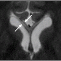
The paired nasal bones are attached to each other and to the nasal spine of the frontal bone. They are grooved on their deep surface by one or more anterior ethmoidal nerves. These vertically orientated grooves can be seen on a radiograph and should not be mistaken for fractures ( Fig. 1.9 ).
Linear lucencies in the nasal bones that run vertically are grooves for the ethmoidal nerves. Horizontally orientated lucencies are likely to be fractures.

- 1.
Frontonasal synchondrosis
- 2.
Nasal spine of frontal bone
- 3.
Groove for anterior ethmoidal nerve
THE BONY ORBIT ( Fig. 1.10 )
The orbit is a four-sided pyramidal bony cavity whose skeleton is contributed to by several bones of the skull. The base of the pyramid is open and points anteriorly to form the orbital rim. Lateral, superior, medial and inferior walls converge posteromedially to an apex , onto which the optic foramen opens, transmitting the optic nerve and ophthalmic artery from the optic canal .

- 1.
Supraorbital notch
- 2.
Nasal septum
- 3.
Infraorbital foramen
- 4.
Superior orbital fi ssure
- 5.
Zygomaticofrontal suture
- 6.
Greater wing of sphenoid
- 7.
Nasolacrimal canal
- 8.
Lateral aspect of inferior orbital fi ssure
- 9.
Orbital process of maxillary bone
The lateral orbital wall is strong and is formed by the zygomatic bone in front and the greater wing of the sphenoid behind. It separates the orbital cavity from the temporal fossa.
The superior wall, or roof, is thin and undulating and separates the orbit from the anterior cranial fossa. It is formed by the orbital plate of the frontal bone in front and the lesser wing of the sphenoid behind.
The medial orbital wall is a thin bone contributed to by maxillary, lacrimal and ethmoid bones, with a small contribution from the sphenoid bone at the apex. It separates the orbit from the nasal cavity, ethmoid air cells and anterior part of sphenoid. The bone between the orbit and ethmoids is paper thin and is known as the lamina papyracea .
Infection can pass easily from ethmoid sinusitis across the paper-thin lamina papyracea into the medial aspect of the orbit.
The inferior wall, or floor, is formed by the orbital process of the maxillary bone, separating the orbit from the cavity of the maxillary sinus. The orbital process of the maxillary bone also extends superomedially to contribute to the medial part of the orbital rim, and the zygoma contributes to the orbital floor laterally. Near the apex there is a tiny bit of palatine bone in the orbital floor.
The orbit has a superolateral depression for the lacrimal gland (see later Fig. 1.24A and B ) and a medial groove for the lacrimal sac and its duct . It also bears the optic foramen, two fissures and a groove in its floor to house the infraorbital nerve.
The superior orbital fissure is a triangular slit between the greater and lesser wings of the sphenoid. Its medial end is wider than its lateral end and is very close to the optic foramen in the apex of the cavity. It transmits the first division of the fifth, and the third, fourth and sixth cranial nerves, as well as the superior ophthalmic veins and a branch of the middle meningeal artery. The middle meningeal artery may communicate with the ophthalmic artery, forming one of the anastomotic connections between internal and external carotid systems.
The inferior orbital fissure is a slit between the lateral and inferior walls of the orbit as they converge on the apex. It runs downward and laterally, and its posteromedial end is close to the medial end of the superior fissure. In its posterior part it forms an opening between the orbit and the pterygopalatine fossa, and more anteriorly it forms an opening between the orbital cavity and the infratemporal fossa. It transmits the infraorbital nerve, which is a branch of the maxillary division of the fifth cranial nerve after it has passed from the middle cranial fossa into the pterygopalatine fossa via the foramen rotundum. It also transmits the infraorbital artery, a branch of the maxillary artery and the inferior ophthalmic veins.
The infraorbital groove runs from the inferior orbital fissure in the floor of the orbit before dipping down to become the infraorbital canal ( Fig. 1.11 ). The nerve emerges from the canal onto the anterior surface of the maxillary bone through the infraorbital foramen.

- 1.
Nasal septum
- 2.
Maxillary sinus
- 3.
Middle nasal turbinate
- 4.
Inferior nasal turbinate
- 5.
Superior meatus
- 6.
Middle meatus
- 7.
Inferior meatus
- 8.
Ethmoid infundibulum
- 9.
Uncinate process
- 10.
Maxillary ostium
- 11.
Maxillary infundibulum
- 12.
Infraorbital nerve
- 13.
Alveolar process of maxilla
- 14.
Ethmoid sinus
- 15.
Sphenoid sinus
- 16.
Sphenoethmoidal recess
- 17.
Superior turbinate
- 18.
Greater palatine canal
The periorbita is a fibrous covering that lines the bony cavity of the orbit. It is continuous with the dura through the optic canal and superior orbital fissure. It closes over the inferior orbital fissure, separating the orbit from the infratemporal and pterygopalatine fossae.
RADIOLOGY OF THE BONY ORBIT
Plain Films
The orbits may be assessed on OF20 and OM projections (see Fig. 1.4 ). Asymmetry between the superior orbital fissures is common.
A straight line is seen running through the orbit from the superolateral part of the rim inferiorly and medially. This is caused by the X-ray beam hitting the curving greater wing of the sphenoid at a tangent and is known as the innominate line .
Computed Tomography
The bony orbit and its soft-tissue contents are demonstrated very well by CT (see Figs. 1.10B and 1.11A ). Axial or coronal images may be obtained. Coronal imaging shows the floor of the orbit and is useful for the assessment of trauma where a fracture is suspected.
Magnetic Resonance Imaging
MRI (see later Fig. 1.23 ) is more valuable for demonstration of the soft-tissue contents of the orbit than the bone.
The Nasal Cavity and Paranasal Sinuses (see Figs. 1.8, 1.11 )
The nasal cavity is a passage from the external nose anteriorly to the nasopharynx posteriorly. The frontal, ethmoid, sphenoid and maxillary sinuses form the paired paranasal sinuses and are situated around, and drain into, the nasal cavity. The entire complex is lined by mucus-secreting epithelium.
THE NASAL CAVITY
This is divided in two by the nasal septum in the sagittal plane. The nasal septum is part bony and part cartilaginous. The floor of the nasal cavity is the roof of the oral cavity and is formed by the palatine process of the maxilla, with the palatine bone posteriorly. The lateral walls of the cavity are formed by contributions from the maxillary, palatine, lacrimal and ethmoid bones. These walls bear three curved extensions known as turbinates or conchae , which divide the cavity into inferior, middle and superior meati , each lying beneath the turbinate of the corresponding name. The space above the superior turbinate is the sphenoethmoidal recess .
- ■
The sphenoid air cells drain into the sphenoethmoidal recess.
- ■
The posterior group of ethmoidal air cells drain into the superior meatus.
- ■
The frontal sinus opens in the most anterior opening of the middle meatus. The anterior ethmoidal air cells and maxillary sinus drain into the middle meatus at the hiatus semilunaris, below the ethmoid bulla.
- ■
The nasolacrimal duct opens into the inferior meatus, draining the lacrimal secretions.
Blood Supply of the Nasal Cavity
The sphenopalatine artery is the terminal part of the maxillary artery. It passes with its associated nerves through the sphenopalatine foramen from the pterygopalatine fossa to the nasal cavity posterior to the superior meatus. It has medial branches to the nasal septum and lateral branches to the lateral wall of the nose and turbinates.
The greater palatine artery supplies some of the lower part of the nasal cavity by branches that pass through the incisive foramen in the anterior part of the hard palate.
The superior labial branch of the facial artery supplies some branches to the anteroinferior part of the nasal septum and the nasal alae.
Anterior and posterior ethmoidal branches of the ophthalmic artery from the internal carotid artery pass through the cribriform plate to supply the superior part of the nasal cavity.
Little’s area is a vascular region of mucosa in the anterior and inferior part of the nasal septum supplied by branches of the sphenopalatine, greater palatine and facial arteries. This is a common site of anterior epistaxis.
THE PARANASAL SINUSES
The Frontal Sinuses
These lie between the inner and outer tables of the frontal bone above the nose and medial part of the orbits; they vary greatly in size and are often asymmetrical. They may extend into the orbital plate of the frontal bone.
The Ethmoid Sinuses
These consist of a labyrinth of bony cavities or cells situated between the medial walls of the orbit and the lateral walls of the upper nasal cavity. Enlargements of anterior cells towards the frontal bone are called agger nasi cells, and enlargements of posterior cells below the apex of the orbit are known as Haller’s cells.
The Sphenoid Sinuses
These paired cavities in the body of the sphenoid are often incompletely separated from each other or may be subdivided further into smaller bony cells. They are so closely related to the ethmoid air cell anteriorly that it may be difficult to distinguish a boundary. The anatomical relationships of the sphenoid sinus are of considerable importance. The sella turcica, bearing the pituitary gland with the optic chiasm anteriorly, is superior. The cavernous sinus and contents run along its lateral walls. The floor of the sphenoid sinus forms the roof of the nasopharynx (see also Chapter 2 Chapter 2 , Pituitary Gland).
The Maxillary Sinuses
The maxillary sinuses, or antra, are the largest of the paranasal sinuses. They are sometimes described as having a body and four processes.
The processes comprise: (1) the orbital process , which extends superomedially to contribute to the medial rim of the orbit; (2) the zygomatic process , which is continuous with the zygomatic arch; (3) the alveolar process , which bears the teeth; and (4) the palatine process , which forms the roof of the mouth and floor of the nasal cavity.
The body of the maxilla is roughly pyramidal in shape, with its apex projecting superomedially between the orbit and nasal cavity. It houses the maxillary sinus. It has an anterior surface that is directed downward and laterally and forms part of the contour of the cheek. It has a curved infratemporal or posterior surface, and this also forms the anterior wall of the infratemporal fossa. Its orbital or superior surface is smooth and triangular and separates the sinus from the orbital cavity. The nasal or medial surface forms the lateral wall of the lower part of the nasal cavity, onto whose middle meatus the sinus drains. The medial wall of the sinus is continued superiorly as a bony projection known as the uncinate process . The maxillary ostium opens superiorly into the infundibulum , which is the channel between the inferomedial aspect of the orbit laterally and the uncinate process medially. The region of the ostium, infundibulum and middle meatus is important clinically and is known as the ostiomeatal complex .
Development of the Paranasal Sinuses in Infants and Children
The maxillary sinuses are the first to appear and are visible radiologically from a few weeks after birth. They continue to grow and develop throughout childhood. The tooth-bearing alveolar process does not begin to develop until the age of 6 years. Full pneumatization of the maxillary sinus is not achieved until there has been complete eruption of the permanent dentition in early adulthood.
The frontal sinuses are not visible on the skull radiograph until the age of 2 years and achieve adult proportions by the age of 14 years. Asymmetry is common, and one or both may fail to develop. Absence of both may be associated with persistence of the metopic suture between the two halves of the frontal bone.
Development of the ethmoids occurs parallel to that of the frontal sinuses.
Pneumatization of the sphenoid sinus commences at 3 years of age and may extend into the greater wings of the sphenoid or clinoid processes. The degree of pneumatization is variable and relevant to trans-sphenoidal hypophysectomy.
During fundoscopic ethmoidal sinus surgery (FESS) the anterior ethmoidal artery is considered unprotected and at risk of injury if there are pneumatized supraorbital anterior ethmoid air cells. The anterior ethmoidal arteries are considered protected if adjacent to the lateral lamella or fovea ethmoidalis in the absence of supraorbital pneumatized anterior ethmoid air cells (see Fig. 1.12A and B ).

RADIOLOGY OF THE NASAL CAVITY AND PARANASAL SINUSES
Computed Tomography (see Figs. 1.8, 1.11 )
CT scanning in axial and, more importantly, coronal planes provides excellent visualization of the paranasal sinuses (see Fig. 1.11 ). Particular attention is paid to the region of the ostiomeatal complex, where the maxillary, frontal and anterior ethmoidal sinuses drain, and the sphenoethmoid recess and superior meatus, into which the sphenoid and posterior ethmoid sinuses drain. The pneumatized sinuses should contain nothing but air.
Magnetic Resonance Imaging
MRI is good at demonstrating the sinuses, as the bony septa, which have no signal themselves, are lined by high-signal mucosa on T 2 scans.
Embolization for epistaxis: When cautery of the bleeding area and nasal packing and other surgical methods fail to control epistaxis, embolization may be successful. Angiographic assessment of the facial, sphenopalatine and greater palatine branches of the external carotid circulation is most likely to identify the source of bleeding. The ethmoidal branches of the ophthalmic artery may also need to be visualized and embolized. In all vessels, the microcatheter to be used for embolization must be advanced distal to branches with a high potential for dangerous anastomotic collaterals, such as the middle meningeal or ophthalmic arteries. Embolization of the superior labial branch of the facial artery may be associated with necrosis of the nasal alae.
The Mandible and Teeth (see Figs. 1.13–1.19 )
THE MANDIBLE (see Fig. 1.13 )
The mandible is composed of two halves united at the symphysis menti . Each half comprises a horizontal body and a vertical ramus joined at the angle of the mandible. The ramus has two superior projections, the coronoid process anteriorly and the condylar process posteriorly, separated by the mandibular (or condylar ) notch . The coronoid process gives attachment to the temporalis muscle, and the condylar process (or head of mandible) articulates with the base of the skull at the temporomandibular joint. The body of the mandible bears the alveolar border with its 16 tooth sockets.

- 1.
Oblique line
- 2.
Mental foramen
- 3.
Mental protuberance
- 4.
Mental tubercle
- 5.
Body of mandible
- 6.
Coronoid process
- 7.
Mandibular notch
- 8.
Condylar process
- 9.
Neck
- 10.
Ramus
- 11.
Angle
- 12.
Mandibular foramen
- 13.
Mylohyoid line


- 1.
Articular tubercle
- 2.
Temporomandibular fossa
- 3.
Head
- 4.
External auditory meatus
- 5.
Neck

- 1.
Mandibular condyle
- 2.
Articular tubercle/eminence
- 3.
Parotid gland
- 4.
External auditory canal
- 5.
Mastoid process of the temporal bone

- 1.
Bone
- 2.
Lamina dura
- 3.
Periodontal membrane
- 4.
Root canal
- 5.
Pulp cavity
- 6.
Dentine
- 7.
Enamel

- 1.
Condylar process of mandible
- 2.
Temporomandibular fossa
- 3.
Hard palate
- 4.
Maxillary sinus
- 5.
Medial wall of maxillary sinus
- 6.
Floor of maxillary sinus
- 7.
Condylar canal
- 8.
Ramus of mandible
- 9.
Angle of mandible
- 10.
Body of mandible
- 11.
Mandibular notch
- 12.
Upper left incisor
- 13.
Hyoid bone (projected laterally)
- 14.
Ear lobe

- 1.
Resorbing roots of a deciduous tooth
- 2.
Unerupted teeth
The mandibular canal runs in the ramus and body of the bone, transmitting the inferior alveolar artery (branch of the maxillary artery) and nerve (branch of the mandibular division of the trigeminal nerve). The mandibular canal opens proximally as the mandibular foramen on the inner surface of the upper ramus, and its distal opening is the mental foramen on the external surface of the body below and between the two premolars.
The muscles of the floor of the mouth, including the medial pterygoid muscles, are attached to the inner surface of the mandible and the muscles of mastication are attached to its outer surface.
THE TEMPOROMANDIBULAR JOINT (see Figs. 1.14–1.16 )
This is a synovial joint between the condyle of the mandible and the temporal bone. The temporal articular surface consists of a fossa posteriorly, the temporomandibular fossa , and a prominence anteriorly, the articular tubercle . The head of the mandible sits in the fossa at rest and glides anteriorly on to the articular tubercle when fully open. The joint is least stable during occlusion.
The articular surfaces are covered with fibrous cartilage. In addition, a fibrocartilaginous disc divides the joint into separate smaller upper and larger lower compartments, each lined by a synovial membrane. The disc is described as having anterior and posterior bands with a thin zone in the middle and is attached to the joint capsule. The anterior band is also attached to the lateral pterygoid muscle. The posterior band is attached to the temporal bone by bands of fibres called the translational zone. No communication between joint compartments is possible unless the disc is damaged. The upper compartment is involved in translational movements and the lower in rotational movements.
THE TEETH: NOMENCLATURE AND ANATOMY (see Figs. 1.17–1.19 )
There are 20 deciduous or milk teeth; in the adult these are replaced by 32 permanent teeth. The complement of teeth in each quadrant is as follows:
- ■
In the child: two incisors, one canine, two molars and
- ■
In the adult: two incisors, one canine, two premolars, three molars
The teeth are referred to by their position in each of the four quadrants. The relevant quadrant is designated by two arms of a cross, and the tooth by its position relative to the midline. The permanent teeth are referred to by number and the milk teeth by capital letter. Thus the second right lower premolar in the adult is designated 5 and the second left upper molar in a child’s milk dentition is designated E.
Each tooth has its own root embedded in a separate socket. The neck of the tooth is covered by the firm fibrous tissue of the gum, and this is covered by the mucous membrane of the mouth. The exposed intraoral part of the tooth is the crown , and this is covered by enamel, which is the hardest and most radio-opaque substance in the body. The remainder of the tooth is composed mostly of dentine, which is of a radiographic density similar to compact bone. A radiolucent pulp cavity occupies the middle of the tooth and is continuous with the root canal , which transmits the nerves and vessels from the supporting bone. The root and neck of the tooth are surrounded by the periodontal membrane , which forms a radiolucent line around them on the radiograph. A dense white line of bone surrounds this and is known as the lamina dura . This surrounds the root of each tooth and is continuous with the lamina dura of the adjacent teeth around the margin of the alveolar crest.
RADIOLOGY OF THE MANDIBLE, TEMPOROMANDIBULAR JOINTS AND TEETH (see Figs. 1.13–1.19 )
Plain Films
The mandible may be seen on skull radiographs and dedicated oblique views of the mandible. The mandibular canal and mandibular and mental foramina may be identified on radiographs of the mandible.
There are also special views for the temporomandibular joints, and a full radiographic study of these includes images of each joint with the mouth open and closed. The teeth can be radiographed on small films, occlusal films, placed close up against them inside the mouth, which provide excellent detail.
The symphysis menti fuses at 2 years of age. Eruption of the milk dentition is normally complete by this time. The permanent dentition develops in the mandible and maxilla during childhood and can be identified radiographically. Most have calcified by 3 years of age. The roots of the milk teeth are resorbed as the permanent teeth erupt. The medial teeth erupt before the lateral teeth, and lower teeth erupt approximately 6–12 months before the upper teeth. The first permanent molar erupts at 6 years of age, and all the permanent dentition is present by the age of 12 or 13 years except the third molar (wisdom tooth), which does not appear until early adulthood.
The mandibular canal and mandibular and mental foramina may be identified on radiographs of the mandible.
Computed Tomography (see Fig. 1.13 )
High-resolution CT is used in the assessment of fractures or bony abnormalities of the mandible.
Magnetic Resonance Imaging (see Fig. 1.16 )
MRI is excellent for the demonstration of the internal temporomandibular joint anatomy. The anterior and posterior bands and the thin zone of the disc are identifiable, as are the disc attachments. Cine MRI studies can also be performed with sagittal images of the joint that show the meniscoligamentous complex in varying degrees of mouth opening and closing.
Dental Pantomography (see Fig. 1.18 )
The dental pantomogram gives a panoramic image of both dental arches, as well as the mandible, temporomandibular joints and lower maxilla. This study is obtained using special equipment that moves around the patient’s face as the radiograph is being taken, mapping out the lower face and jaw in a straight line.
The Orbital Contents ( Figs. 1.20–1.24 )
The orbit contains the globe, the extraocular muscles (including levator palpebrae), the lacrimal gland, the optic nerve and the ophthalmic vessels. The whole is embedded in fat. The orbit is limited anteriorly by the orbital septum . This is a thin layer of fascia that extends from the orbital rim to the superior and inferior tarsal plates, separating the orbital contents from the eyelids. A fascial layer, the periorbita , lines the bony cavity of the orbit and this is continuous with the dura mater of the brain through the superior orbital fissure and optic canal.


- 1.
Anterior chamber
- 2.
Iris
- 3.
Anterior aspect of lens
- 4.
Posterior aspect of lens
- 5.
Vitreous body
- 1.
Vitreous body
- 2.
Retinal surface
- 3.
Optic nerve
- 4.
Retrobulbar fat
- 5.
Lateral rectus muscle
- 6.
Lateral wall of bony orbit

- 1.
Nasal septum
- 2.
Nasal bone
- 3.
Ethmoid air cells
- 4.
Globe of left eye
- 5.
Sclera
- 6.
Optic nerve
- 7.
Medial rectus muscle
- 8.
Lateral rectus muscle
- 9.
Superior ophthalmic vein
- 10.
Optic canal
- 11.
Anterior clinoid process (pneumatized)
- 12.
Superior orbital fi ssure
- 13.
Middle cranial fossa
- 14.
Greater wing of sphenoid
- 15.
Frontal process of zygomatic bone
- 16.
Temporal fossa/temporalis muscle

- 1.
Optic nerve
- 2.
Sclera
- 3.
Globe
- 4.
Lacrimal gland
- 5.
Lateral rectus muscle
- 6.
Medial rectus muscle
- 7.
Optic canal
- 8.
Temporal lobe
- 9.
Greater wing of sphenoid
- 10.
Frontal process of zygomatic bone
- 11.
Temporalis muscle
- 1.
Superior rectus muscle
- 2.
Inferior rectus muscle
- 3.
Medial rectus muscle
- 4.
Lateral rectus muscle
- 5.
Superior oblique muscle
- 6.
Lacrimal gland
- 7.
Frontal bone
- 8.
Crista galli
- 9.
Infra-orbital foramen/nerve
- 10.
Roof of orbit
Related posts:
Stay updated, free articles. Join our Telegram channel

Full access? Get Clinical Tree


