In the pediatric age group, primary cardiac tumors are extremely rare; a prevalence of 0.0017 to 0.28% in autopsy series has been reported. However, with the increasing use of CT and MRI, they may be identified more often, and radiologists should be familiar with their imaging findings.
The majority of primary cardiac tumors are benign, and less then 10% are malignant. However, the majority of cases will be secondary tumors and/or metastatic disease.
Table 1.80 Cardiac tumors—septal/myocardial tumor
Diagnosis
Findings
Comments
Benign tumors
Fibroma
Solitary tumor.
Primary location ventricular septum.
Calcifications are common.
Low attenuation on CT.
Isointense on T1-weighted and hypointense on T2-weighted images. Little to no enhancement after gadolinium.
Can extend into the ventricular conduction system and cause arrhythmia.
Can be associated with Gorlin syndrome.
Teratoma
Atrial and ventricular wall.
Heterogeneous, encapsulated cystic masses.
Rare tumor, most often arising from the pericardial space.
Hemangioma
Can be found anywhere in the heart.
Subendocardial nodules (2–4 cm diameter).
Exceedingly rare cardiac tumor.
Histiocytic nodule
Nodular deposits on the valves or ventricular endocardium on US.
Also known as infantile histiocytic cardiomyopathy, oncocytic cardiomyopathy, histiocytic cardiomyopathy, Purkinje cell tumor, focal lipid cardiomyopathy, and idiopathic infantile cardiomyopathy.
Malignant tumors
Intrapericardial pheochromocytoma
Vascular tumor arising from the autonomic paraganglia.
Predominantly in atrial septum.
Metastatic cardiac tumors
Multiple discrete firm epicardial nodules on autopsy.
Melanoma has a tendency to spread to the heart.
Estimated to be 20 times more common than primary cardiac tumors.
Table 1.81 Cardiac tumors—intracardiac tumor
Diagnosis
Findings
Comments
Benign tumors
Rhabdomyoma
Bright intramural mass on cardiac US.
Predominantly in the ventricular myocardium.
Often multiple lesions.
Can protrude into the cardiac cavity.
Most common benign primary cardiac tumor (> 60%). Preferential cardiac location intraventricular. Strongly related to tuberous sclerosis, present in 43%–72% of patients.
Myxoma
Primary location in left atrium (90%), but also found in right atrium.
Mostly a solitary tumor.
Pediculated mass with irregular nonhomogeneous small lucencies on US.
Heterogeneous mass with low attenuation on CT. Heterogeneous mass, bright on T2-weighted imaging with heterogeneous enhancement after gadolinium.
Most common primary cardiac tumor in adults.
Can be associated with Carney complex (autosomal dominant syndrome of cardiac myxomas and hyperpigmented skin lesions), LAMB (l entigines, a trial myxoma, m ucocutaneous myxoma, b lue nevi), and NAME (n aevi, a trial myxoma, m yxoid neurofibroma, e pithelides).
Hemangioma
Can be found anywhere in the heart.
Subendocardial nodules (2–4 cm diameter).
Exceedingly rare cardiac tumor.
Malignant tumors
Angiosarcoma
Most common malignant tumor in adults.
Primary location right atrium.
Broad-based mass with epicardial, endocardial, and/or intracavitary extension.
Areas of high signal intensity on T1-weighted images, focal or linear along pericardium.
Can be seen as an extension of tumor thrombus (e.g., Wilms tumor).
Hematologic spread most common.
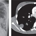
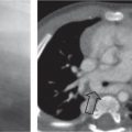
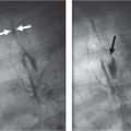
Fig. 1.213a, b Rhabdomyosarcoma. (a) Axial T1-weighted post-contrast. Note the presence of lung metastases (arrowhead). Biopsy proved primary, maliganant cardiac rhabdomyosarcoma in atrial septum (arrow). (Courtesy of A. Taylor, MD, FRCP, FRCR, Cardio-respiratory Unit, UCL Institute of Child Health & Great Ormond Street Hospital for Children, London, United Kingdom.) (b) Balanced steady-state free precession cine image (systolic frame) of the same patient shows extension into the anterior leaflet of the mitral valve (arrow), which is a finding suggestive of malignancy.Fig. 1.214 Metastatic osteosarcoma to the right ventricle in a 17-year-old girl (arrow).
Only gold members can continue reading. Log In or Register to continue