Projection on radiograph defines the valve replacement.
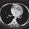
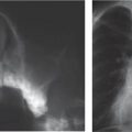
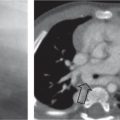
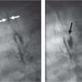
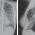
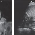
Fig. 1.227a, b Coarctation aorta. (a) An 18-year-old man with coarctation aorta after endovascular stent treatment (arrow). (b) Three-dimensional SSD of the endovascular stent.Fig. 1.228a–c Melody stent. (a) A 14-year-old girl after Melody stent placement (arrow). (b) Melody stent. (c) Melody stent with broken strut (see inset).Fig. 1.229 A 16-year-old boy after endovascular treatment of ASD using an Amplatzer device (see inset).Fig. 1.230a–c Cardiac arrhythmia. (a) A 9-year-old girl with tetralogy of Fallot. An epicardial pacemaker has been placed. Note the presence of a Melody valve replacement. (b) A 15-year-old girl with an implantable cardioverter defibrillator; the lead is positioned within the right ventricle. (c) A 1-year-old boy with a subcutaneous implantable cardioverter defibrillator in combination with electrocardial pacing.Fig. 1.231 A 17-year-old boy with a mitral valve replacement.Fig. 1.232 Schematic representation of valve replacements on the anteroposterior/posteroanterior chest radiograph (left) and lateral radiograph (right).
P
pulmonary
A
aortic
T
tricuspid
M
mitral
Further Reading
Ablin DS, Azouz EM, Jain KA. Large intrathoracic tumors in children: imaging findings. AJR Am J Roentgenol 1995;165:925–934Andrén-Sandberg A, Dervenis C. Pancreatic pseudocysts in the 21st century. Part I: classification, pathophysiology, anatomic considerations and treatment. JOP 2004;5:8–24Andronikou S, Wieselthaler N. Modern imaging of tuberculosis in children: thoracic, central nervous system and abdominal tuberculosis. Pediatr Radiol 2004;34(11):861–875Agrons GA, Courtney SE, Stocker JT, Markowitz RI. From the archives of the AFIP: lung disease in premature neonates: radiologic-pathologic correlation. Radiographics 2005;25(4):1047–1073Backer CL, Mavroudis C. Congenital Heart Surgery Nomenclature and Database Project: vascular rings, tracheal stenosis, pectus excavatum. Ann Thorac Surg 2000;69(4 Suppl):S308–318Beall DP, Ly J, Bell JP, et al. Pediatric extraskeletal osteosarcoma. Pediatr Radiol 2008;38:579–582Berdon WE. Rings, slings, and other things: vascular compression of the infant trachea updated from the midcentury to the millennium—the legacy of Robert E. Gross, MD, and Edward B. D. Neuhauser, MD. Radiology 2000;216(3):624–632Bining HJ, Saigal G, Chankowsky J. Kingella kingae spondylodiscitis in a child. Br J Radiol 2006;79(947):e181–183Brien EW, Mirra JM, Ippolito V, Vaughan L. Clear-cell chondrosarcoma with elevated alkaline phosphatase, mistaken for osteosarcoma on biopsy. Skeletal Radiol 1996;25:770–774Brill PW, Winchester P, Kleinman PK. Differential diagnosis I: diseases simulating abuse. In: Kleinman PK, ed. Diagnostic imaging of child abuse. 2nd ed. St. Louis: Mosby, 1998:178–196Calder AD, Owens CM. Computed tomography of the central and peripheral airways. In: Donoghue VP, ed. Radiological imaging of the neonatal chest. Berlin: Springer-Verlag Berlin and Heidelberg GmbH & Co, 2007:177–195Carden KA, Boiselle PM, Waltz DA, Ernst A. Tracheomalacia and tracheobronchomalacia in children and adults: an in-depth review. Chest 2005;127(3):984–1005Castellote A, Enriquez G, Lucaya J. Congenital malformations of the chest beyond the neonatal period. In Carty H, Brunelle F, Stringer DA, Kao SC-S, eds. Imaging children. Oxford, UK: Churchill Livingstone, 2005:1049–1074Chao S, Mullins ME, Slanetz PJ. Posterior mediastinal pheochromocytoma. AJR Am J Roentgenol 2001;176:1408Cheema JI, Grissom LE, Harcke HT. Radiographic characteristics of lowerextremity bowing in children. Radiographics 2003;23:871–880Chernick V, Boat TF, Wilmott RW, Bush A. Kendig’s disorders of the respiratory tract in children. Philadelphia: Elsevier Inc., 2006Cleveland RH. A radiologic update on medical diseases of the newborn chest. Pediatr Radiol 1995;25(8):631–637Cohen MM Jr. A comprehensive and critical assessment of overgrowth and overgrowth syndromes. Adv Hum Genet 1989;18:181–303, 373–376Dahnert W. Radiology review manual. Philadelphia: Lippicott Williams and Wilkins, 2007Diren HB, Kutluk MT, Karabent A, Göçmen A, Adalioĝlu G, Kenanoĝlu A. Primary hypertrophic osteoarthropathy. Pediatr Radiol 1986; 16:231–234Dodd GD 3rd, Ledesma-Medina J, Baron RL, Fuhrman CR. Posttrans-plant lymphoproliferative disorder: intrathoracic manifestations. Radiology 1992;184(1):65–69Donnelly LF. Practical issues concerning imaging of pulmonary infection in children. J Thorac Imaging 2001;16(4):238–250Donnelly LF, Frush DP. Langerhans cell histiocytosis showing low-attenuation mediastinal mass and cystic lung sisease. AJR 2000; 174:877–878Donnelly LF, Klosterman LA. The yield of CT of children who have complicated pneumonia and noncontributory chest radiography. AJR Am J Roentgenol 1998;170(6):1627–1631Donoghue VP. Hyaline membrane disease and its complications. In: Donoghue VP, ed. Radiological imaging of the neonatal chest. Berlin: Springer-Verlag Berlin and Heidelberg GmbH & Co, 2007:67–79Donoghue VP. Transient tachypneoa of the newborn. In: Donoghue VP, ed. Radiological imaging of the neonatal chest. Berlin: Springer-Verlag Berlin and Heidelberg GmbH & Co, 2007:81–83Ebel KD, Blickman H, Willich E, Richter E, eds. The mediastinum. In: Differential diagnosis in pediatric radiology. Stuttgart: Thieme, 1999:155–200Edwards DK 3rd, Berry CC, Hilton SW. Trisomy 21 in newborn infants: chest radiographic diagnosis. Radiology 1988;167:317–318Effmann EL, Merten DF, Kirks DR, Pratt PC, Spock A. Adult respiratory distress syndrome in children. Radiology 1985;157(1):69–74Effman EL. Chest wall. In Kuhn JP, Slovis TL, Haller JO, Caffey J, eds. Caffey’s pediatric diagnostic imaging. Philadelphia: Elsevier Inc., 2004:817–856Effman EL, Kuhn JP. Lungs and airways. In Kuhn JP, Slovis TL, Haller JO, Caffey J, eds. Caffey’s pediatric diagnostic imaging. Philadelphia: Elsevier Inc., 2004:891–1072Elliott M, Roebuck D, Noctor C, et al. The management of congenital tracheal stenosis. Int J Pediatr Otorhinolaryngol 2003;67 Suppl 1:S183–192Feigin RD. Textbook of pediatric infectious diseases. 5th ed. Philadelphia: Saunders, 2004Garcés-Iñigo EF, Leung R, Sebire NJ, McHugh K. Extrarenal rhabdoid tumours outside the central nervous system in infancy. Pediatr Radiol 2009;39:817–822Gardner DJ, Azouz EM. Solitary lucent epiphyseal lesions in children. Skeletal Radiol 1988;17:497–504Gartner L, Pearce CJ, Saifuddin A. The role of the plain radiograph in the characterisation of soft tissue tumours. Skeletal Radiol 2009;38:549–558Geier A, Lammert F, Gartung C, Nguyen HN, Wildberger JE, Matern S. Magnetic resonance imaging and magnetic resonance cholangiopancreaticography for diagnosis and pre-interventional evaluation of a fluid thoracic mass. Eur J Gastroenterol Hepatol 2003; 15:429–431Gilsanz V, Perez FJ, Campbell PP, Dorey FJ, Lee DC, Wren TA. Quantitative CT reference values for vertebral trabecular bone density in. Radiology 2009;250:222–227Giron J, Fajadet P, Sans N, et al. Diagnostic approach to mediastinal masses. Eur J Radiol 1998;27(1):21–42Gladish GW, Sabloff BM, Munden RF, Truong MT, Erasmus JJ, Chasen MH. Primary thoracic sarcomas. Radiographics 2002;22:621–637Glass RB, Norton KI, Mitre SA, Kang E. Pediatric ribs: a spectrum of abnormalities. Radiographics 2002;22:87–104Goldfarb CA, Manske PR, Busa R, Mills J, Carter P, Ezaki M. Upper-extremity phocomelia reexamined: a longitudinal dysplasia. J Bone Joint Surg Am 2005;87:2639–2648Goldman AB, Kaye JJ. Macrodystrophia lipomatosa: radiographic diagnosis. AJR Am J Roentgenol 1977;128:101–105Grayev AM, Boal DK, Wallach DM, Segal LS. Metaphyseal fractures mimicking abuse during treatment for clubfoot. Pediatr Radiol 2001;31:559–563Green DM, Breslow NE, Beckwith JB, Norkool P. Screening of children with hemihypertrophy, aniridia, and Beckwith-Wiedemann syndrome in patients with Wilms tumor: a report from the National Wilms Tumor Study. Med Pediatr Oncol 1993;21:188–192Greenspan A, Jundt G, Remagen W. Differential diagnosis in orthopaedic oncology. 2nd ed. Philadelphia: Lippincott Williams & Wilkins, 2007Heller GD, Haller JO, Berdon WE, Sane S, Kleinman PK. Punctate thymic calcification in infants with untreated Langerhans’ cell histiocytosis: report of four new cases. Pediatr Radiol 1999;29(11):813–815Hernanz-Schulman M. Vascular rings: a practical approach to imaging diagnosis. Pediatr Radiol 2005;35(10):961–979Houser JR, Kan JH. Langerhans cell histiocytosis of the epiphysis. Pediatr Radiol 2008;38:85518Howling SJ, Northway WH Jr, Hansell DM, Moss RB, Ward S, Muller NL. Pulmonary sequelae of bronchopulmonary dysplasia survivors: high-resolution CT findings. AJR Am J Roentgenol 2000;174(5):1323–1326Hussmann J, Russell RC, Kucan JO, Khardori R, Steinau HU. Soft-tissue calcifications: differential diagnosis and therapeutic approaches. Ann Plast Surg 1995;34:138–147Jaggers J, Balsara K. Mediastinal masses in children. Semin Thorac Cardiovasc Surg 2004;16(3):201–208Jaramillo D, Shapiro F, Hoffer FA, et al. Posttraumatic growth-plate abnormalities: MR imaging of bony-bridge formation in rabbits. Radiology 1990;175:767–773Jeanes AC, Owens CM. Chest imaging in the immunocompromised child. Paediatr Respir Rev 2002;3(1):59–69Jeung MY, Gangi A, Gasser B, et al. Imaging of chest wall disorders. Radiographics 1999;19:617–637Jeung MY, Gasser B, Gangi A, et al. Imaging of cystic masses of the mediastinum. Radiographics 2002;22:S79–93John SD, Ramanathan J, Swischuk LE. Spectrum of clinical and radio-graphic findings in pediatric mycoplasma pneumonia. Radiographics 2001;21(1):121–131Jolles H, Henry DA, Robertson JR, Cole TJ, Spratt JA. Mediastinitis following median sternotomy: CT findings. Radiology 1996; 201:463–466Keller KA, Barnes PD. Rickets vs. abuse: a national and international epidemic. Pediatr Radiol 2008;38:1210–1216Khanna G, Sato TS, Ferguson P. Imaging of chronic recurrent multifocal osteomyelitis. Radiographics 2009;29:1159–1177Kilic D, Tercan F, Sahin E, Blien A, Hatipoglu A. Unusual radiological manifestations of the echinococcus infection in the thorax. J Thorac Imaging 2006;21(1):32–36Kimonis VE, Mehta SG, Digiovanna JJ, Bale SJ, Pastakia B. Radiological features in 82 patients with nevoid basal cell carcinoma (NBCC or Gorlin) syndrome. Genet Med 2004;6:495–502Klein DM, Barbera C, Gray ST, Spero CR, Perrier G, Teicher JL. Sensitivity of objective parameters in the diagnosis of pediatric septic hips. Clin Orthop Relat Res 1997;338:153–159Kleinman PK. Diagnostic imaging of child abuse. 2nd ed. St. Louis: Mosby, 1989Kleinman PK. Problems in the diagnosis of metaphyseal fractures. Pediatr Radiol 2008;38 Suppl 3:S388–394Kleinman PK. Skeletal trauma: general considerations. In: Kleinman PK, ed. Diagnostic imaging of child abuse. 2nd ed. St. Louis: Mosby, 1998:168–177Ko SF, Hsieh MJ, Ng SH, et al. Imaging spectrum of Castleman’s disease. AJR Am J Roentgenol 2004;182:769–775Koh DM, Hansell DM. Computed tomography of diffuse interstitial lung disease in children. Clin Radiol 2000;55(9):659–667Kothari NA, Kramer SS. Bronchial diseases and lung aeration in children. J Thorac Imaging 2001;16(4):207–223Kozlowski K, Sutcliffe J, Barylak A, et al. Hypophosphatasia. Review of 24 cases. Pediatr Radiol 1976;5:103–117Kuhn JP. Diaphragm. In Kuhn JP, Slovis TL, Haller JO, Caffey J, eds. Caffey’s pediatric diagnostic imaging. Philadelphia: Elsevier Inc., 2004:857–866Kuhn JP. Mediastinum. In Kuhn JP, Slovis TL, Haller JO, Caffey J, eds. Caffey’s pediatric diagnostic imaging. Philadelphia: Elsevier Inc., 2004:1160–1224Kuhn JP. Pleura. In Kuhn JP, Slovis TL, Haller JO, Caffey J, eds. Caffey’s pediatric diagnostic imaging. Philadelphia: Elsevier Inc., 2004:867–890Kumar R, Madewell JE, Swischuk LE, Lindell MM, David R. The clavicle: normal and abnormal. Radiographics 1989;9:677–706Lachman R. Taybi and Lachman’s radiology of syndromes, metabolic disorders and skeletal dysplasias. 5th ed. St. Louis: Mosby, 2006Laor T, Jaramillo D. MR imaging insights into skeletal maturation: what is normal? Radiology 2009;250:28–38Le Goffc, Cormier-Daire V. Genetic and molecular aspects of acromelic dysplasia. Pediatr Endocrinol Rev 2009;6:418–423Levesque M, Legmann P, Le Cloirec A, Deybach JC, Nordmann Y. Radiological features in congenital erythropoietic porphyria (Gunther’s disease). Report of 3 cases. Pediatr Radiol 1988;18:62–66Levine MS, Borden S 4th, Gill FM. Sternal cupping: a new finding in childhood sickle cell anemia. Radiology 1982;142:367–370Makley JT, Dunn MJ. Prostaglandin synthesis by osteoid osteoma. Lancet 1982;2:42Mar WA, Taljanovic MS, Bagatell R, et al. Update on imaging and treatment of Ewing sarcoma family tumors: what the radiologist needs to know. J Comput Assist Tomogr 2008;32:108–118McCahon E. Lung tumours in children. Paediatr Respir Rev 2006; 7(3):191–196McCarville MB, Kaste SC, Pappo AS. Soft-tissue malignancies in infancy. AJR Am J Roentgenol 1999;173:973–977McHugh K. Mediastinal and chest tumours. In Carty H, Brunelle F, Stringer DA, Kao SC-S, eds. Imaging children. Oxford, UK: Churchill Livingstone, 2005:1147–1172McPhillips M. Infection. In Carty H, Brunelle F, Stringer DA, Kao SC-S, eds. Imaging children. Oxford, UK: Churchill Livingstone, 2005:1075–1118McQueen FM. Magnetic resonance imaging in early inflammatory arthritis: what is its role? Rheumatology (Oxford) 2000;39:700–706Mehta AV, Chidambaram B, Suchedina AA, Garrett AR. Radiologic abnormalities of the sternum in Turner’s syndrome. Chest 1993; 104:1795–1799Merten DF. Diagnostic imaging of mediastinal masses in children. AJR Am J Roentgenol 1992;158(4):825–832Moppett J, Oakhill A, Duncan AW. Second malignancies in children: the usual suspects? Eur J Radiol 2001;38:235–248Mora S, Gilsanz V. Establishment of peak bone mass. Endocrinol Metab Clin North Am 2003;32:39–63Muecke EC, Currarino G. Congenital widening of the pubic symphysis: associated clinical disorders and roentgen anatomy of affected bony pelves. Am J Roentgenol Radium Ther Nucl Med 1968; 103:179–185Newman B. Congenital bronchopulmonary foregut malformations: concepts and controversies. Pediatr Radiol 2006;36(8):773–791Nguyen ML, Jones NF. Undergrowth: brachydactyly. Hand Clin 2009; 25:247–255O’Connor JF, Cohen J. Dating fractures. In: Kleinman PK, ed. Diagnostic imaging of child abuse. 2nd ed. St. Louis: Mosby, 1998:168–177Oestreich AE. The lateral clavicle hook-an acquired as well as a congenital anomaly. Pediatr Radiol 1981;11:147–150Offiah A, van Rijn RR, Perez-Rossello JM, Kleinman PK. Skeletal imaging of child abuse (non-accidental injury). Pediatr Radiol 2009; 39:461–470OMIM—Online Mendelian Inheritance in Man. Available at http://www-ncbi-nlm-nih-gov.easyaccess1.lib.cuhk.edu.hk/omimOlsen OE, Owens CM. Diffuse interstitial lung disease. In Carty H, Brunelle F, Stringer DA, Kao SC-S, eds. Imaging children. Oxford, UK: Churchill Livingstone, 2005:1119–1132Oner Y, Uzun M, Tokgöz N, Tali ET. Isolated true anterior thoracic meningocele. AJNR Am J Neuroradiol 2004;25(10):1828–1830Owens CM. Radiology of diffuse interstitial pulmonary disease in children. Eur Radiol 2004;14 Suppl 4:L2–12Owens CM. Meconium aspiration. In: Donoghue VP, ed. Radiological imaging of the neonatal chest. Berlin: Springer-Verlag Berlin and Heidelberg GmbH & Co, 2007:85–97Paajanen H, Hermunen H, Karonen J. Pubic magnetic resonance imaging findings in surgically and conservatively treated athletes with osteitis pubis compared to asymptomatic athletes during heavy training. Am J Sports Med 2008;36:117–121Patel MD, Filly RA. Homozygous achondroplasia: US distinction between homozygous, heterozygous, and unaffected fetuses in the second trimester. Radiology 1995;196:541–545Pilling DW, Pilling P. The neonatal chest. In Carty H, Brunelle F, Stringer DA, Kao SC-S, eds. Imaging children. Oxford, UK: Churchill Livingstone, 2005:1023–1048Poznanski AK, Fernbach SK, Berry TE. Bone changes from prostaglandin therapy. Skeletal Radiol 1985;14:20–25Prosser I, Maguire S, Harrison SK, Mann M, Sibert JR, Kemp AM. How old is this fracture? Radiologic dating of fractures in children: a systematic review. AJR Am J Roentgenol 2005;184:1282–1286Resnick D, Greenway G. Distal femoral cortical defects, irregularities, and excavations. Radiology 1982;143:345–354Restrepo CS, Martinez S, Lemos DF, et al. Imaging appearances of the sternum and sternoclavicular joints. Radiographics 2009;29:839–859Rossi UG, Owens CM. The radiology of chronic lung disease in children. Arch Dis Child 2005;90(6):601–607Ryan S. Postnatal imaging of chest malformations. In: Donoghue VP, ed. Radiological imaging of the neonatal chest. Berlin: Springer-Verlag Berlin and Heidelberg GmbH & Co, 2007:139–162Shabshin N, Schweitzer ME, Morrison WB, Carrino JA, Keller MS, Gris-som LE. High-signal T2 changes of the bone marrow of the foot and ankle in children: red marrow or traumatic changes? Pediatr Radiol 2006;36:670–676Simmons BP, Southmayd WW, Riseborough EJ. Congenital radioulnar synostosis. J Hand Surg Am 1983;8:829–838Slovis TL, Chapman S. Evaluating the data concerning vitamin D insufficiency/deficiency and child abuse. Pediatr Radiol 2008;38:1221–1224Spranger J. Radiologic nosology of bone dysplasias. Am J Med Genet 1989;34:96–104Steinberg ME, Steinberg DR. Classification systems for osteonecrosis: an overview. Orthop Clin North Am 2004;35:273–283, vii-viiiSubbarao K. Periosteal reactions in pediatrics. Indian J Pediatr 1987;54:45–52Temtamy SA, Aglan MS. Brachydactyly. Orphanet J Rare Dis 2008; 3:15 Ulkü R, Eren N, Cakir O, Balci A, Onat S. Extrapulmonary intrathoracic hydatid cysts. Can J Surg 2004;47(2):95–98Van Rijn RR, Wilde JC, Bras J, Oldenburger F, McHugh KM, Merks JH. Imaging findings in noncraniofacial childhood rhabdomyosarcoma. Pediatr Radiol 2008;38:617–634Varich LJ, Laor T, Jaramillo D. Normal maturation of the distal femoral epiphyseal cartilage: age-related changes at MR imaging. Radiology 2000;214:705–709Wahlgren H, Mortensson W, Eriksson M, Finkel Y, Forsgren M, Leinonen M. Radiological findings in children with acute pneumonia: age more important than infectious agent. Acta Radiol 2005;46(4):431–436Waters PM, Smith GR, Jaramillo D. Glenohumeral deformity secondary to brachial plexus birth palsy. J Bone Joint Surg Am 1998;80:668–677Wetherell RG, Amis AA, Heatley FW. Measurement of acetabular erosion. The effect of pelvic rotation on common landmarks. J Bone Joint Surg Br 1989;71:447–451Whitten CR, Khan S, Munneke GJ, Grubnic S. A diagnostic approach to mediastinal abnormalities. Radiographics 2007;27(3):657–671Williams HJ, Alton HM. Imaging of paediatric mediastinal abnormalities. Pediatr Respir Rev 2003;4(1):55–66Wong KS, Chiu CH, Huang YC, Lin TY. Childhood and adolescent tuberculosis in northern Taiwan: an institutional experience during 1994–1999. Acta Paediatr 2001;90:943–947Worthy SA, Flint JD, Muller NL. Pulmonary complications after bone marrow transplantation: high-resolution CT and pathologic findings. Radiographics 1997;17(6):1359–1371Yekeler E, Tunaci M, Tunaci A, Dursun M, Acunas G. Frequency of sternal variations and anomalies evaluated by MDCT. AJR Am J Roentgenol 2006;186:956–960Zook PD, Winter TC 3rd, Nyberg DA. Iliac angle as a marker for Down syndrome in second-trimester fetuses: CT measurements. Radiology 1999;211:447–451Zubler V, Mengiardi B, Pfirrmann CW, et al. Bone marrow changes on STIR MR images of asymptomatic feet and ankles. Eur Radiol 2007;17:3066–3072
Only gold members can continue reading. Log In or Register to continue