CHAPTER 26 Hemangioma
Epidemiology
Along with schwannomas, hemangiomas are the most common tumors of the facial nerve canal; 0.7% of all petrous temporal bone tumors are hemangiomas. They can occur at any age, but usually are found in adults. Within the temporal bone, the most common location of a hemangioma is the geniculate fossa (region of first genu of the facial nerve canal in the region of the geniculate ganglion), with the internal auditory canal being a less common location for these tumors. Rarely are hemangiomas found in the second genu (facial nerve recess) region.
Clinical Features
As this lesion arises from perineural (facial nerve) vasculature, it is not surprising that it usually presents with facial nerve dysfunction. Symptoms may be due to nerve compression or frank invasion. Symptom onset may be relatively acute (arising over weeks) or more insidious. Symptoms are dependent on the location of the lesion; geniculate fossa lesions present with isolated facial nerve dysfunction, whereas internal auditory canal lesions present with both facial nerve dysfunction and sensorineural hearing loss due to the proximity of the vestibulocochlear (eighth cranial) nerve to the facial nerve in the internal auditory canal.
Pathology
Hemangiomas are slow-growing benign vascular tumors arising from perineural vessels that surround the intratemporal facial nerve. As these vessels appear to be most numerous along the geniculate and proximal tympanic portions of the facial nerve, it is not surprising that those locations are where hemangiomas most commonly occur. Hemangiomas may be divided into three histologic subtypes: (1) ossifying hemangioma (associated with spicules of lamellar bone), (2) cavernous hemangioma (associated with large vascular channels), and (3) capillary hemangioma (associated with small vascular channels). It should be noted that all three histologic subtypes may be present in a single lesion. Hemangiomas can cause symptoms both by nerve compression and invasion. They can invade the nerve (even when lesions are quite small), requiring resection of that part of the nerve for definitive treatment.
Treatment
Complete surgical resection of a hemangioma is curative. No chemotherapy or radiation therapy is necessary. When a lesion is small, it may still be extraneural (causing symptoms by nerve compression only) and the lesion may be completely resected with preservation of seventh nerve function. However, whether large or small, if the lesion has invaded the seventh nerve, that portion of the nerve along with the hemangioma may need to be resected with placement of a nerve graft. Such patients who require grafting have greater morbidity than those who undergo nerve-sparing procedures.
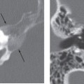
Imaging Findings
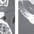
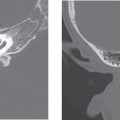
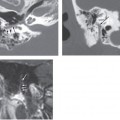
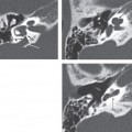

Stay updated, free articles. Join our Telegram channel

Full access? Get Clinical Tree


