Chapter 190
Hemangiomas
Epidemiology
Hemangiomas are proliferative endothelial vascular lesions that are identified by their characteristic clinical appearance and course. These lesions may arise in the buccal space. Forty percent of hemangiomas are present at birth and 60% appear within the first few months of life. Hemangiomas are more common in females than in males (5:1). Common sites of occurrence include skin, face, orbits, larynx, nasal cavity, and deep neck spaces.
Clinical Findings
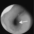
Superficial hemangiomas are bright red papular lesions. Subcutaneous hemangiomas often present as a bluish mass that may be difficult to differentiate from a venous malformation or arteriovenous malformation.
Hemangiomas rapidly grow during the first 12 to 18 months of life (proliferative phase). This is followed by gradual regression (involuting phase) over the next 6 to 10 years. Approximately half of all lesions will completely involute whereas the remainder will partially involute. Incomplete involution may result in residual telangiectasias, hypoplastic patches, or scarring. The majority of hemangiomas have an uneventful course, with spontaneous and complete involution. More advanced lesions may cause severe facial disfigurement.
Pathology
The proliferative phase consists of proliferating plump endothelial cells with frequent mitoses. The end of the proliferative phase is characterized by a reduction in the mitotic activity, progressive flattening of the cells, and an abundance of mast cells. Apoptosis and progressive endothelial cells encompassed by large ectatic vascular channels in a matrix of fibrofatty tissue characterize the involutional phase.
Treatment