Hematoma/Seroma
KEY FACTS
Imaging
 Quite well-defined heterogeneous mass ± echogenic pseudosolid appearance
 Multilaminated whorled appearance
 Anechoic cystic areas
 Echogenic foci due to gas locules
 Surrounding soft tissue edema
 Mild surrounding hyperemia
 No internal hyperemia
 Underlying cause of hematoma, such as muscle tear or vascular malformation, may be apparent
IMAGING
General Features
Ultrasonographic Findings
 Appearances depend on chronicity
 Acute: Fairly well-defined heterogeneous mass in subcutaneous or muscle layer
 Subacute and chronic: Well-defined, variable echogenicity depending on liquefaction and degree of reparative organization
 Organizing hematoma
 Hemophiliac pseudotumor
 Rectus sheath hematoma


 anterior to the patella
  . The hematoma shows a heterogeneous, hypoechoic, pseudosolid appearance. A more fluid component is present at the superficial aspect of the hematoma
  .
.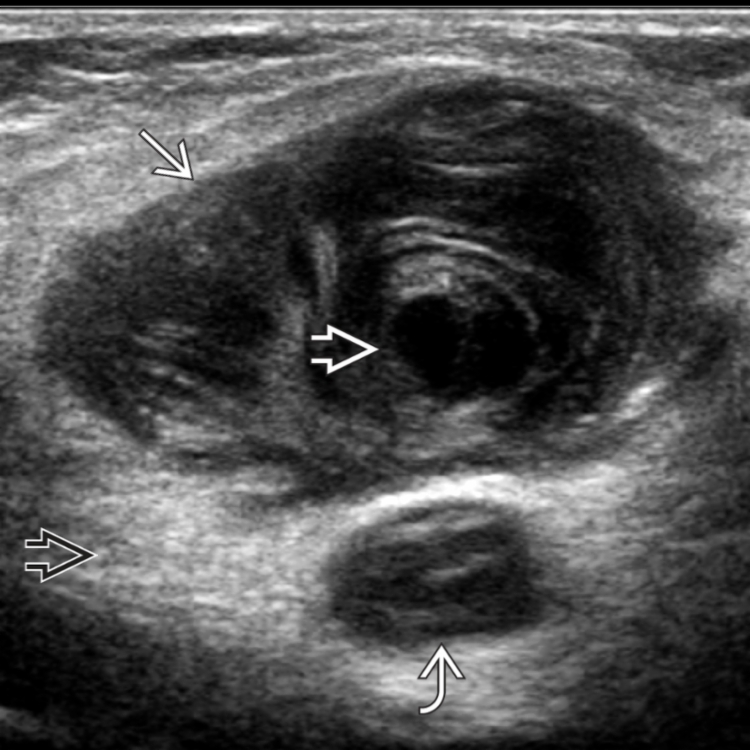
 . It shows typical multilaminated whorled appearance with posterior enhancement and a central anechoic
  fluid component. Another smaller hematoma
  is present posteriorly. A moderate degree of surrounding edema
  is present.
 .
.


