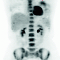
Fig. 16.1
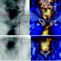
MIP image demonstrates the persistence of high metabolism of the hepatic lesion in the fifth segment. The other two secondary nodules do not appear in MIP reconstruction because they are subcentimetric and also have only a limited increase in carbohydrate consumption, and their activity is masked by the background activity of the liver and the physiological renal excretion
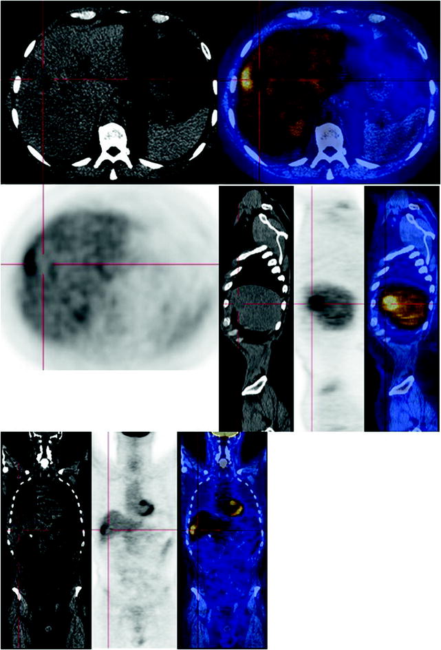
Fig. 16.2
 Laryngeal Squamous Carcinoma: Staging
 Radio-Treated Cancer of the Posterior Hemi-Circumference of the Anal Canal: Post-Actinic Fibrosis
 Urothelial Carcinoma: Follow-Up After Surgery
 Lymphocytic Interstitial Pneumonia in Patient with History of Breast Cancer
 Metastatic Breast Carcinoma: Restaging After Neoadjuvant Chemotherapy
 Bone Metastases from Breast Cancer: Progression of Disease and Subsequent Response to Radiotherapy


The CT scan shows a subcapsular, uneven mass in the hepatic V segment, with markedly hypodense internal component due to colliquation
Related posts:
Laryngeal Squamous Carcinoma: Staging
Radio-Treated Cancer of the Posterior Hemi-Circumference of the Anal Canal: Post-Actinic Fibrosis
Urothelial Carcinoma: Follow-Up After Surgery
Lymphocytic Interstitial Pneumonia in Patient with History of Breast Cancer
Metastatic Breast Carcinoma: Restaging After Neoadjuvant Chemotherapy
Bone Metastases from Breast Cancer: Progression of Disease and Subsequent Response to Radiotherapy
Stay updated, free articles. Join our Telegram channel
Full access? Get Clinical Tree
