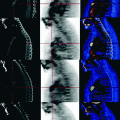
Fig. 13.1
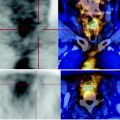
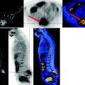
Sagittal CT-PET reconstruction: in correspondence of the rectal anastomosis, at the level of the metal clips, solid, inhomogeneous tissue which has no pathological glucose consumption can be seen. This element is attributable to post-surgical scarring
13.4 Conclusions
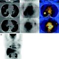
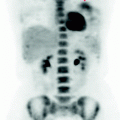
The PET scan today shows two metastatic liver nodules with high carbohydrate metabolism, showing recurrence of disease.
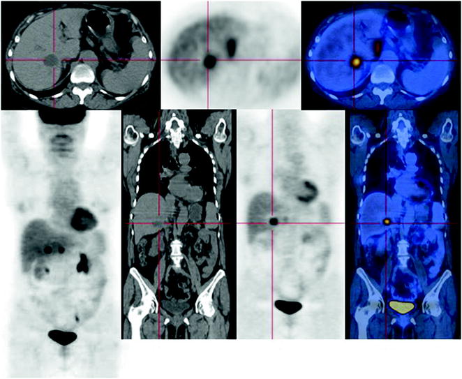
Fig. 13.2


PET scan shows two hepatic nodules, respectively at the VI and II segment that have high consumption of glucose. CT scan identifies only that the sixth segment
Stay updated, free articles. Join our Telegram channel

Full access? Get Clinical Tree


