Diagnosis |
Findings |
Comments |
Variations in normal anatomy |
Sternal foramen, sclerotic bands, manubriosternal, and sternoxiphoidal fusions. |
Cortical irregularity, expansion, and soft-tissue mass help distinguish a pathologic process from normal variations in anatomy. |
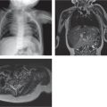
Pectus excavatum |
Concave posterior depression of the sternum often with rotation along the transverse plane results in a reduction of the prevertebral space, leftward displacement and axial rotation of the heart, and reduction in the space occupied by the lungs (usually the left lung). |
Most common congenital deformity of the sternum. The pectus (Haller) index, derived by dividing the transverse diameter of the chest by the anteroposterior (AP) diameter, obtained on axial computed tomography (CT) or magnetic resonance imaging (MRI). A pectus index greater than 3.25 necessitates surgical correction. |
Trisomy 21 |
Multiple manubrial ossification centers (hypersegmentation). |
Probability of trisomy 21 increases with the number of anomalies present on the chest radiograph (11 rib pairs and a bell-shaped chest). |
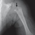
Sickle cell
Fig. 5.1 |
Sternal cupping at the ends of the segments. |
Similar appearance as end plate depressions in vertebral bodies. May be seen in up to 8% of patients. |
Turner syndrome |
Short, premature fusion of manubriosternal junction or mesosternum, decreased ratio of sternal body to manubrium, two ossification centers, bowing, mild pectus excavatum. |
(see Table 5.30 ) |
Poland syndrome |
Deformity of the sternum associated with absence of muscles and clavicle. |
(see Table 5.15 ) |
Chronic recurrent multifocal osteomyelitis (CRMO) |
Hyperostosis and osteomyelitis. |
Chronic inflammatory condition of unknown etiology. Usually multifocal. Children 5–15 y old. Accompanied by synovitis, acne, pustulosis. Adult form is SAPHO (s ynovitis, a cne, p ustulosis, h yperostosis, and o steitis). |