, Marilyn J. Siegel2, Tomasz Miszalski-Jamka3, 4 and Robert Pelberg1
(1)
The Christ Hospital Heart and Vascular Center of Greater Cincinnati, The Lindner Center for Research and Education, Cincinnati, OH, USA
(2)
Mallinckrodt Institute of Radiology, Washington University School of Medicine, St. Louis, Missouri, USA
(3)
Department of Clinical Radiology and Imaging Diagnostics, 4th Military Hospital, Wrocław, Poland
(4)
Center for Diagnosis Prevention and Telemedicine, John Paul II Hospital, Kraków, Poland
Abstract
“Heterotaxy” is simply defined as an abnormality of situs where the internal thoracic and abdominal organs show an arrangement other than situs solitus or inversus [1]. The term is synonymous with “situs ambiguous.”
“Heterotaxy” is simply defined as an abnormality of situs where the internal thoracic and abdominal organs show an arrangement other than situs solitus or inversus [1]. The term is synonymous with “situs ambiguous.”
The incidence of heterotaxy is estimated to be 1 in 10,000 live births [2]. It is associated with a spectrum of cardiac malformations varying from relatively simple anomalies to very complex defects [3].
Heterotaxy is not one specific finding but rather it is a constellation of atrial, visceral, and cardiovascular abnormalities. The morphologic spectrum of the heterotaxy syndrome includes atrial isomerism, abnormal thoracic and abdominal visceral situs (in particular the presence or absence of the spleen), anomalous systemic or pulmonary venous return, and associated cardiac defects [4–6].
20.1 Atrial Isomerism
The atria maintain their laterality throughout development; hence, they define the cardiac situs. In normal individuals, the morphologic right atrium is on the same side as the morphologic right-sided thoracic and visceral structures. Likewise, the morphologic left atrium is on the same side as the morphologic left-sided thoracic and abdominal structures.
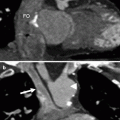
At imaging, the atria can be recognized by their different anatomy (described in detail in Sect. 1.2). Briefly, the right atrium is the more trabeculated atrium with the broad-based appendage and receives blood from the vena cava. The left atrium has a smaller, narrower appendage and receives blood from the pulmonary veins (Fig. 20.1).
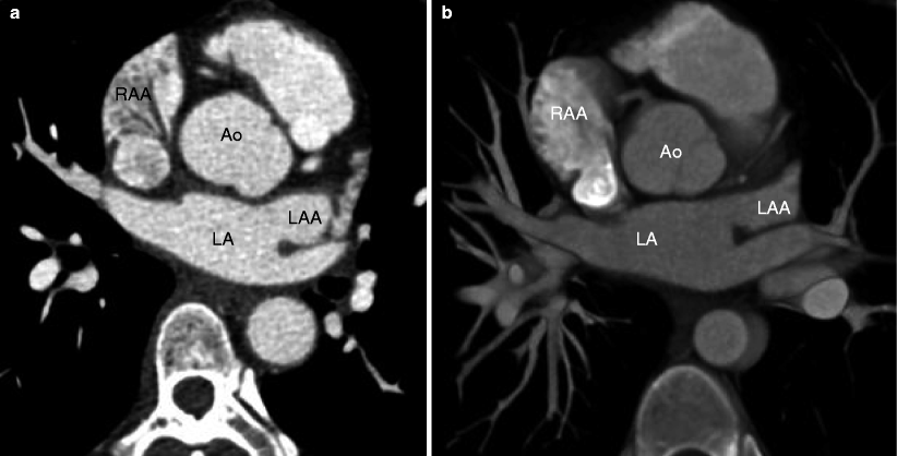
Fig. 20.1
Atrial appendage morphology. Panels (a) and (b) are two axial computed tomography images. The right atrial appendage (RAA) is a blunt, broad-based structure lined with pectinate muscles that form a trabeculated surface. The left atrial appendage (LAA) is smaller and narrower and has a more restricted junction with the left atrium (LA). Ao aorta
Atrial isomerism refers to hearts with bilaterally right or bilaterally left atrial appendages [4–6]. Right atrial isomerism is characterized by the presence of a morphologic right atrium on both the right and left sides of the midline (Fig. 20.2). Likewise, left atrial isomerism is characterized by morphologically bilateral left atria (Fig. 20.3). Although atrial isomerism is often present in heterotaxy syndrome, it is not a reliable term to describe the syndrome because it inaccurately reflects the full constellation of abnormalities.
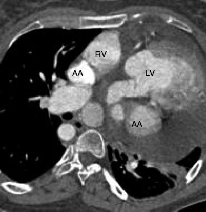
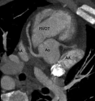
Fig. 20.2
Right atrial isomerism. The atrial appendages (AA) bilaterally are broad-based structures, consistent with right atrial morphology. RV right ventricle, LV left ventricle
Fig. 20.3
Left atrial isomerism. The atrial appendages (AA) bilaterally are elongated and fingerlike, consistent with left atrial morphology. RVOT right ventricular outflow tract
If the atria are not distinguishable based on morphology, then determination of atrial situs can be made based on localization of noncardiac structures, namely, the tracheobronchial tree morphology (i.e., tracheobronchial situs). The right superior branch of the right main bronchus is eparterial, meaning that it is located above the segmental pulmonary artery that supplies the superior lobe of the right lung. The left superior bronchus is hyparterial and passes below the pulmonary arteries. The right, middle, and inferior lobar bronchi and left inferior lobar bronchus are also hyparterial, passing below the arteries. In right isomerism both bronchi are eparterial (Fig. 20.4), and in left isomerism, both bronchi are hyparterial (Fig. 20.5).
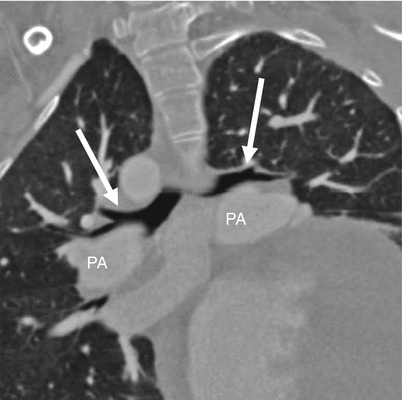
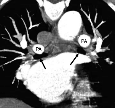
Fig. 20.4
Right isomerism. Both bronchi (arrows) are eparterial and course above the pulmonary arteries (PA)
Fig. 20.5
Left isomerism. Both bronchi (arrows) are hyparterial and course below the pulmonary arteries (PA)
Of note, neither atrial nor tracheobronchial tree morphology is a reliable marker of the position of the ventricles, the cardiac apex, or the great arteries which must be identified and reported separately. A systematic approach to the diagnosis of visceroatrial situs, which is helpful in predicting the likelihood of associated heart disease, is discussed in more detail in Chap. 5.
20.2 Thoracic and Abdominal Situs
There are three types of situs: situs solitus (S), inversus (I), and ambiguous (A). As noted above, heterotaxy is synonymous with situs ambiguous.
Situs solitus is the usual arrangement of the atria and viscera. Situs solitus is characterized by a morphologic right atrium on the right side of the spine, a trilobed right lung with eparterial bronchus, and bilobed left lung with hyparterial bronchus, right-sided liver, left-sided spleen, and right-sided inferior vena cava. The normal ventricular relationship is a D-ventricular loop, with the anatomic right ventricle positioned to the right of the left ventricle. The cardiac apex and stomach are on the left side of the spine (Fig. 20.6).
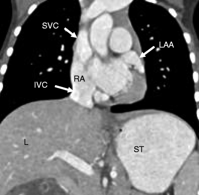
Fig. 20.6
Situs solitus. This is the usual arrangement of organs and vessels. The morphologic right atrium (RA), superior vena cava (SVC) and inferior vena cava (IVC), and liver (L) are on the right side of the thorax. The left atrial appendage (LAA), cardiac apex, and stomach (ST) are on the left
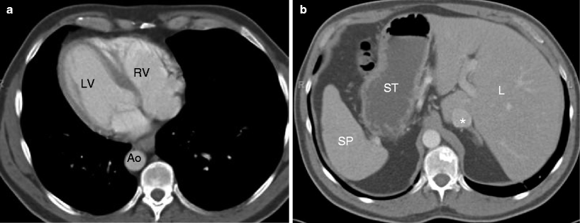
Situs inversus (also called situs inversus totalis) is the “mirror image” of situs solitus. Situs inversus is characterized by a two-lobed right lung with a hyparterial bronchus and three-lobed left lung with eparterial bronchus (i.e., the morphologic left lung is right sided and the morphologic right lung is left sided), left-sided liver, and right-sided spleen. There is usually an L-ventricular loop with the anatomic right ventricle positioned to the left of the left ventricle. The cardiac apex and stomach are right sided (Figs. 20.7 and 20.8).
Get Clinical Tree app for offline access
Fig. 20.7
Situs inversus. The anatomic arrangement is the mirror image of situs solitus. Panel (a


Related posts:


Stay updated, free articles. Join our Telegram channel
Full access? Get Clinical Tree
