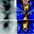
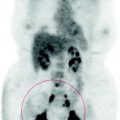
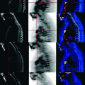
Fig. 79.1
The PET scan shows bone, lung and lymph node secondary disease due to disseminated urothelial carcinoma
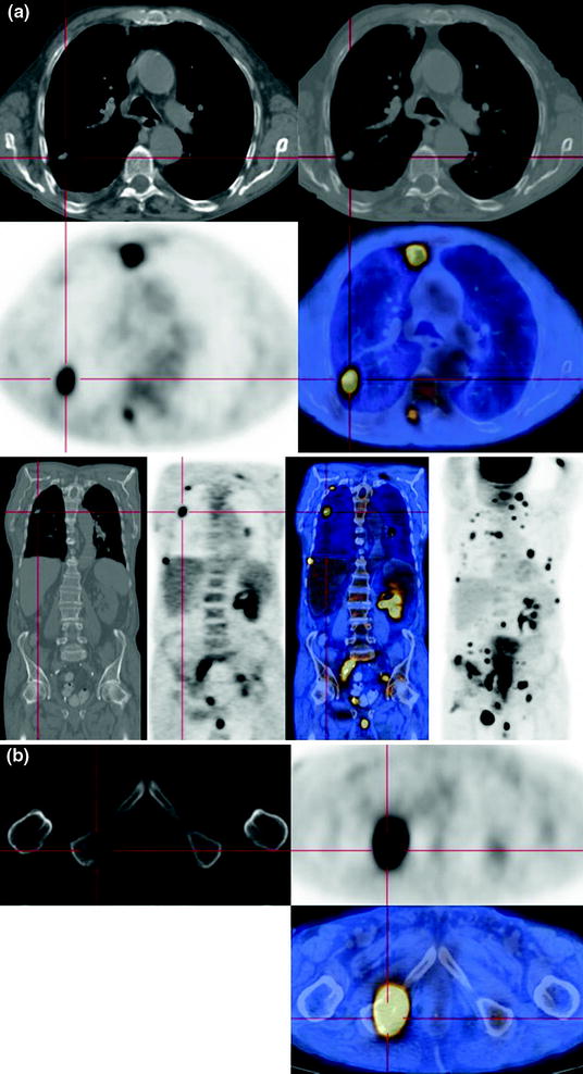
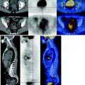
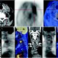
Fig. 79.2
The CT-PET shows multiple pulmonary solid metastatic nodules (a); only two have high concentration of FDG, the other subcentimetric nodules are below the resolving power of the technique. Presence of numerous metastatic bone lesions with extensive metabolism, most of which are not seen on CT because they have not yet determined enough demineralization and destruction, an element that occurs only belatedly (b)
79.5 Key Points
Prostate cancer with normal PSA after surgery rarely develops lytic bone metastases with a high metabolism of glucose. It is clear that the skeletal, lung, and lymph nodal lesions are secondary to the high-grade urothelial neoplasm.
Stay updated, free articles. Join our Telegram channel

Full access? Get Clinical Tree


